
Stock image Right Ventricle


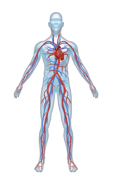
Simple Vector Illustration Of The Circulatory System Focused On The Heart And Lungs With The Names Of Each Part Written In English On A Black Background.
Vector, 0.98MB, 4800 × 3600 eps


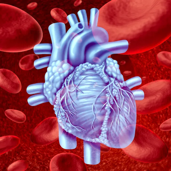
Human Heart Concept Anatomy On A Grunge Background As A Medical Health Care Symbol Or Cardiology Icon Of An Inner Cardiovascular Organ In A 3D Illustration Style.
Image, 13.86MB, 5305 × 3393 jpg

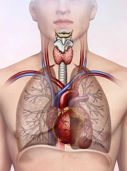
Anemic Hypoxia, Hypoxic, Stagnant, Histotoxic, Arterial PO2 And O2, Arterio-venous Shunts, Respiratory System, 3d Render
Image, 4.05MB, 3840 × 2160 jpg

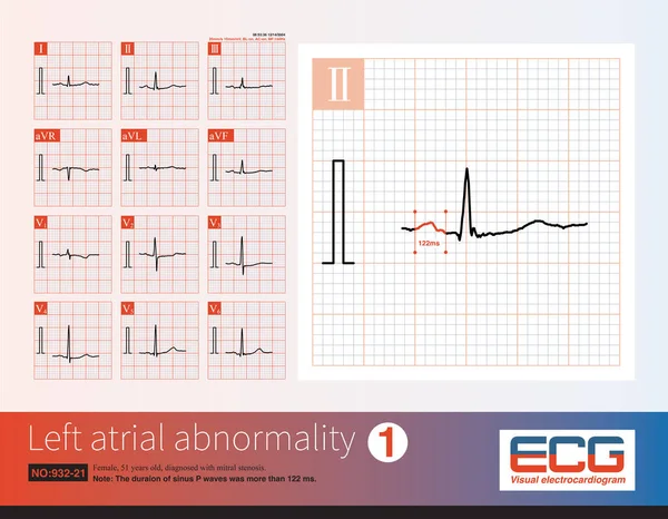
Female, 51 Years Old, Diagnosed With Mitral Stenosis. When This ECG Was Taken, The Patient Still Maintained Sinus Rhythm.Note That The P Wave Duration Was Widened.
Image, 14.21MB, 10000 × 7772 jpg
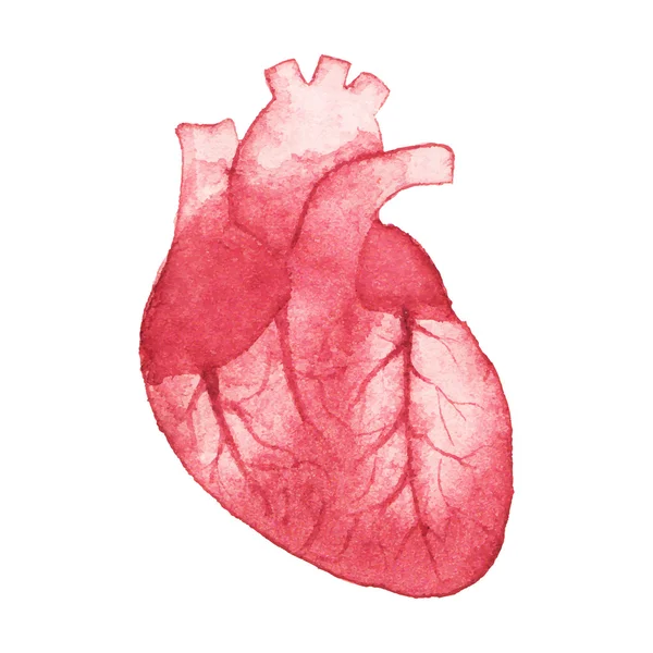
Watercolor Realistic Human Heart On The White Background, Aquarelle. Vector Illustration. Hand-drawn Decorative Element Useful For Invitations, Scrapbooking, Design.
Vector, 10.31MB, 5000 × 5000 eps


Heart Disease Awareness And Cardiovascular Illness Research And Stroke Prevention As A Medical Health Symbol For High Blood Pressure Hypertension And Arrhythmia With 3D Illustration Elements
Image, 7.56MB, 7655 × 3500 jpg
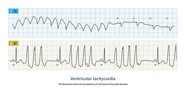
The Illustration Shows The Two Patterns Of Ventricular Tachycardia Episodes.The Green Circle Represents Sinus Rhythm. Picture A Shows Paroxysmal Episodes Of Ventricular Tachycardia, And Picture B Shows Short Bursts.
Image, 10.72MB, 10000 × 5059 jpg

Anti Valentines Day Card, Skeleton Holding A Flower Rose, Vector Illustration
Vector, 4.39MB, 4167 × 4707 eps


Watercolor Seamless Pattern With Realistic Human Heart On The White Background, Aquarelle. Vector Illustration.
Vector, 12.35MB, 5002 × 5003 eps

Anti Valentines Day Banner, My Heart Beats For You. Vector Illustration
Vector, 15.79MB, 3751 × 6542 eps


Viral Myocarditis Or Virus Infection Of The Human Heart Resulting In Inflammation Of The Cardiac Circulatory Organ With 3D Illustration Elements.
Image, 20.08MB, 7085 × 3506 jpg


This Is A Pathological Photo Of Human Left Ventricular Hypertrophy, Showing An Increase In Myocardial Diameter And Interstitial Distance.Magnify 40x.
Image, 42.86MB, 8500 × 8500 jpg
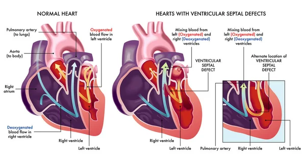
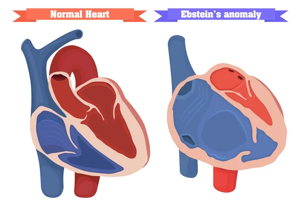
Medical Illustration That Compares A Normal Heart With Hearts Afflicted By Ventricular Septal Defects, An Abnormal Opening (hole) In The Heart, With Annotations.
Vector, 9.41MB, 7000 × 3686 eps
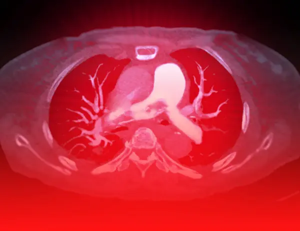
A CTA Pulmonary Artery Reveals A Detailed View Of The Lung Blood Vessels, Capturing The Presence Of A Pulmonary Embolism, A Condition Where A Blood Clot Disrupts Normal Blood Flow.
Image, 2.47MB, 3856 × 2976 jpg

The Typical ST-T Changing Of Left Ventricular Hypertrophy Are: ST Segment Slightly Convex With Downward Sloping Depression; Fusion Of ST Segment And Inverted T Wave; Asymmetry Of Inverted T Wave.
Image, 11.52MB, 10000 × 8453 jpg

Male, 65 Years Old, Was Clinically Diagnosed With Acute Anterior Myocardial Infarction. The Patient Was Treated With A Coronary Stent, But No Reperfusion T Wave Occurred On Day 2.
Image, 19.72MB, 10000 × 8695 jpg

The Presence Of Atrioventricular Dissociation In Wide-complex Tachycardia Is Highly Suggestive Of Ventricular Tachycardia.
Image, 11.09MB, 10000 × 5896 jpg
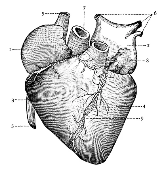
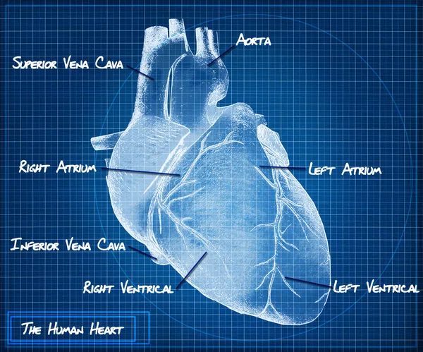
A Typical Representation Of The Section Of The Stomach, With The Parts, 1: Right Auricle; 2: Left Auricle; 3: Right Ventricle; 4: Left Ventricle; 5: Systemic Veins; 6: Pulmonary Veins; 7: Aorta; And Other, Vintage Line Drawing Or Engraving Illustrati
Vector, 5.42MB, 9491 × 9718 eps
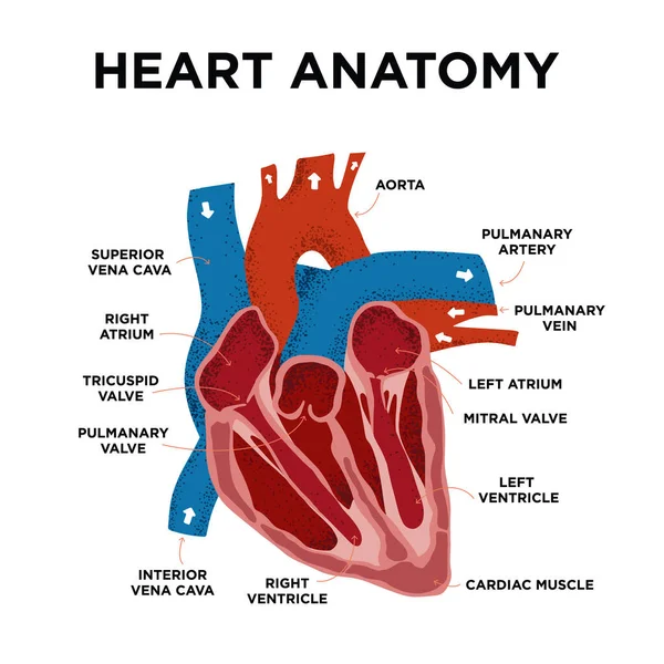
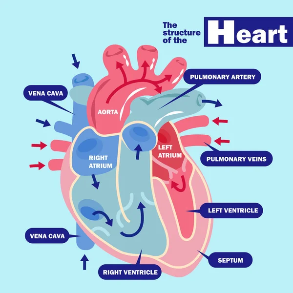
Heart Anatomy Diagram. Human Heart Structure. Labelled Heart Half In Doodle Style. Part Of Heart Foe Education. Hand Drew Vector Illustration.
Vector, 9.06MB, 5000 × 5000 eps
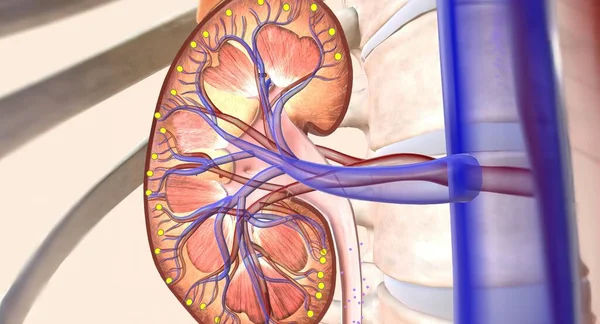
A Synthetic Graft Is An Artificial Tube That Allows The Oxygenated Blood To Flowfrom The Heart To The Rest Of The Body. 3D Rendering
Image, 3.17MB, 7258 × 3920 jpg

A CTA Pulmonary Artery Reveals A Detailed View Of The Lung Blood Vessels, Capturing The Presence Of A Pulmonary Embolism, A Condition Where A Blood Clot Disrupts Normal Blood Flow.
Image, 3.49MB, 5352 × 3229 jpg

In Acute Left Main Occlusion, The Left Ventricular Myocardium Is Massively Ischemic And Necrotic, The Excitatory Potential Of The Left Ventricle Is Weakened, And The Axis May Deviate To The Right .
Image, 12.47MB, 10000 × 6364 jpg

Male, 65 Years Old, Was Clinically Diagnosed With Acute Anterior Myocardial Infarction. The Patient Was Treated With A Coronary Stent, But No Reperfusion T Wave Occurred On Day 2.
Image, 25.24MB, 17956 × 5906 jpg
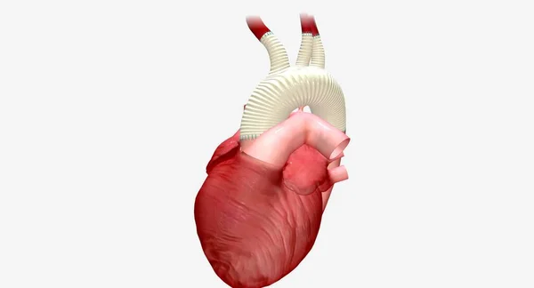
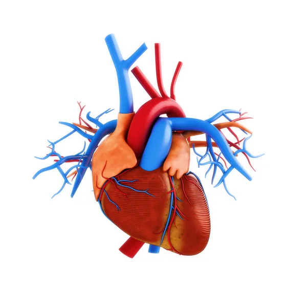
Human Heart Anatomy From A Healthy Body Isolated On White Background As A Medical Health Care Symbol Of An Inner Cardiovascular Organ In A 3D Illustration Style.
Image, 4.29MB, 6188 × 3300 jpg
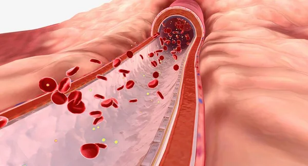
Lipids Are Types Of Fat That Travel Through The Bloodstream. 3D Render
Image, 15.08MB, 7258 × 3920 jpg
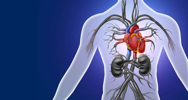
Angiotensin Receptor Neprilysin Inhibitors (ARNIs) Are A Combination Medication That Work By Helping The Kidney Move Extra Sodium Into The Ureter, Where It Is Flushed Away With Urine. 3D Rendering
Image, 14.49MB, 7258 × 3920 jpg

The Doctor Uses The Image As An Educational Tool, Simplifying The Concept Of Pulmonary Embolism To Ensure Patients Comprehend The Diagnosis.Clipping Path.
Image, 3.15MB, 3904 × 3117 jpg
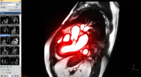
MRI Heart Or Cardiac MRI Magnetic Resonance Imaging Of Heart In Sagittal View Showing Cross-sections Of The Left And Right Ventricle For Detecting Heart Disease On Blurred Monitor.
Image, 3.72MB, 7536 × 4147 jpg
Page 1 >> Next