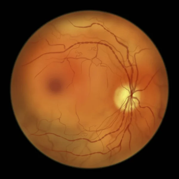
Stock image Diabetic macular edema (DME). Diabetic retinopathy, illustration showing macula edema, abnormal finding on fundoscopic examination of the eye retina in diabetes mellitus

Published: May.16, 2022 07:28:20
Author: katerynakon
Views: 12
Downloads: 1
File type: image / jpg
File size: 2.72 MB
Orginal size: 5000 x 5000 px
Available sizes:
Level: silver
Similar stock images

Non-proliferative Diabetic Retinopathy, Illustration Showing Small Retinal Haemorrhages, Ophthalmoscope View
5000 × 5000