
Stock image Macula Edema

Proliferative Diabetic Retinopathy, Illustration Showing Neovascularization In The Disk And Macula Edema. Abnormal Finding On Fundoscopic Examination Of The Eye Retina In Diabetes Mellitus
Image, 2.85MB, 5000 × 5000 jpg
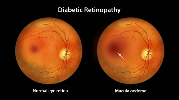
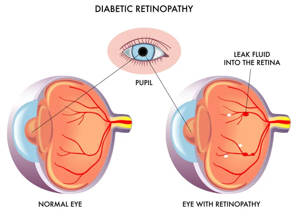
Diabetic Macular Edema (DME), Illustration Showing Normal Eye Retina And Retina With Macula Edema. Fundoscopic Examination Of The Eye Retina In Diabetes Mellitus
Image, 6.24MB, 11738 × 6603 jpg
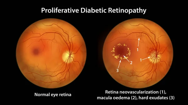
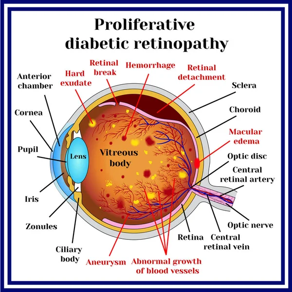
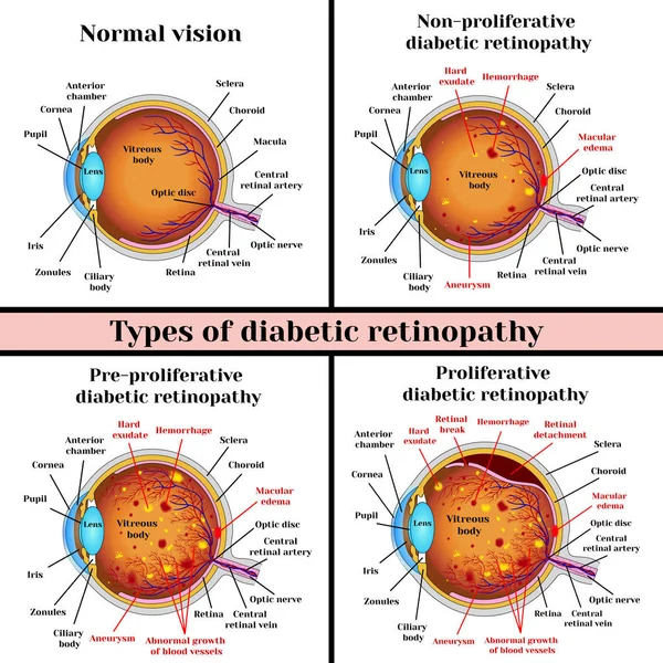
Proliferative Diabetic Retinopathy, Illustration Showing Neovascularization In The Disk And Other Sites, Macula Edema And Hard Exudates. Fundoscopic Examination Of The Eye Retina In Diabetes Mellitus
Image, 6.95MB, 11738 × 6603 jpg
Proliferative Diabetic Retinopathy, Illustration Showing Neovascularization In The Disk And Other Sites, And Macula Edema. Fundoscopic Examination Of The Eye Retina In Diabetes Mellitus
Image, 2.9MB, 5000 × 5000 jpg

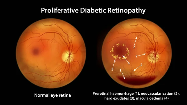
Proliferative Diabetic Retinopathy, Illustration Showing Neovascularization In The Disk And Other Sites, Macula Edema And Hard Exudates. Fundoscopic Examination Of The Eye Retina In Diabetes Mellitus
Image, 3.05MB, 5000 × 5000 jpg

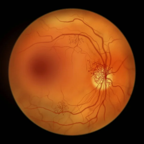
Diabetic Macular Edema (DME). Diabetic Retinopathy, Illustration Showing Macula Edema, Abnormal Finding On Fundoscopic Examination Of The Eye Retina In Diabetes Mellitus
Image, 2.72MB, 5000 × 5000 jpg

Diabetic Retinopathy, 3D Illustration Showing Macula Edema, Optic Disk Edema And Hard Exudates, Abnormal Finding On Fundoscopic Examination Of The Eye Retina In Diabetes Mellitus
Image, 26.67MB, 11738 × 6603 jpg

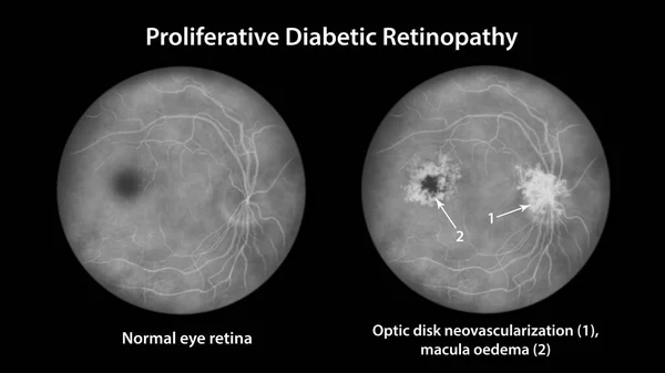
Proliferative Diabetic Retinopathy, Illustration Showing Neovascularization In The Disk And Other Sites, And Macula Edema. Eye Retina In Diabetes Mellitus, Fluorescein Angiography
Image, 2.67MB, 5000 × 5000 jpg
Proliferative Diabetic Retinopathy, Illustration Showing Neovascularization In The Disk And Cystoid Macula Edema. Abnormal Finding On Fundoscopic Examination Of The Eye Retina In Diabetes Mellitus
Image, 6.18MB, 11738 × 6603 jpg
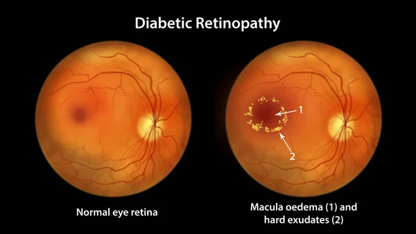
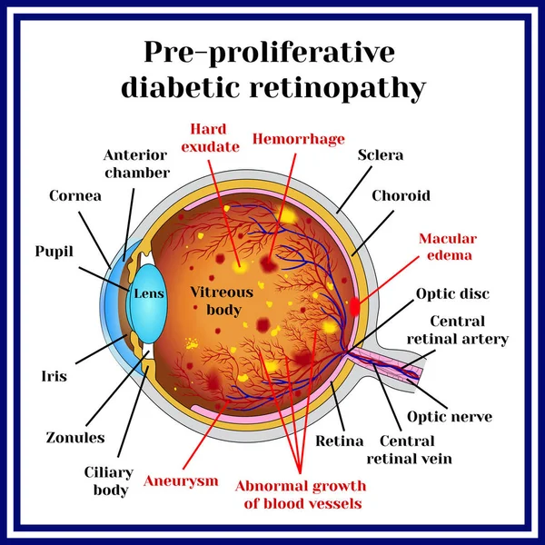
Diabetic Retinopathy, Illustration Showing Macula Edema And Hard Exudates, Abnormal Finding On Fundoscopic Examination Of The Eye Retina In Diabetes Mellitus
Image, 6.56MB, 11738 × 6603 jpg

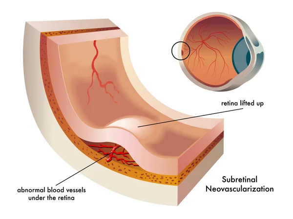
Proliferative Diabetic Retinopathy, 3D Illustration Showing Preretinal Haemorrhage As Horizontal Blood Level, Neovascularization In The Disk And Other Sites, Macula Edema And Hard Exudates
Image, 22.98MB, 11738 × 6603 jpg
Proliferative Diabetic Retinopathy, Illustration Showing Preretinal Haemorrhage As Horizontal Blood Level, Neovascularization In The Disk And Other Sites, Macula Edema And Hard Exudates
Image, 7.07MB, 11738 × 6603 jpg
OCT Of The Eye Reveals Faint Epimacular Membrane And Full Thickness Macular Hole Involving The Fovea, Surrounding Diffuse Macular Oedema Showing Few Cystoid Changes For Follow Up, Selective Focus
Image, 11.49MB, 4800 × 3840 jpg
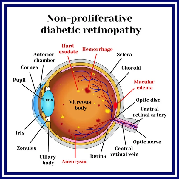

Diabetic Retinopathy. Retinal Damage. Cross Section Of Human Eye. Diabetes. Close-up Of A Macula, Optic Disc, Choroid, Retina, Sclera, And Fovea. Medical Condition. Microaneurysm Of The Small Blood Vessels. Vector Poster
Vector, 5.75MB, 6008 × 3000 eps


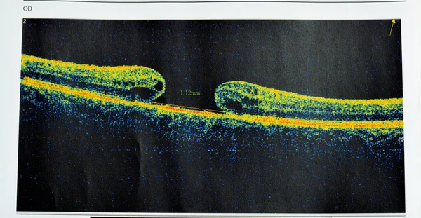
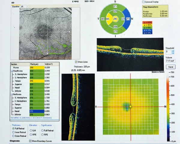
OCT Of The Eye Reveals Faint Epimacular Membrane And Full Thickness Macular Hole Involving The Fovea, Surrounding Diffuse Macular Oedema Showing Few Cystoid Changes For Follow Up, Selective Focus
Image, 12.52MB, 6000 × 3114 jpg

OCT Of The Eye Reveals Faint Epimacular Membrane And Full Thickness Macular Hole Involving The Fovea, Surrounding Diffuse Macular Oedema Showing Few Cystoid Changes For Follow Up, Selective Focus
Image, 13.79MB, 5541 × 3822 jpg
Page 1 >> Next