
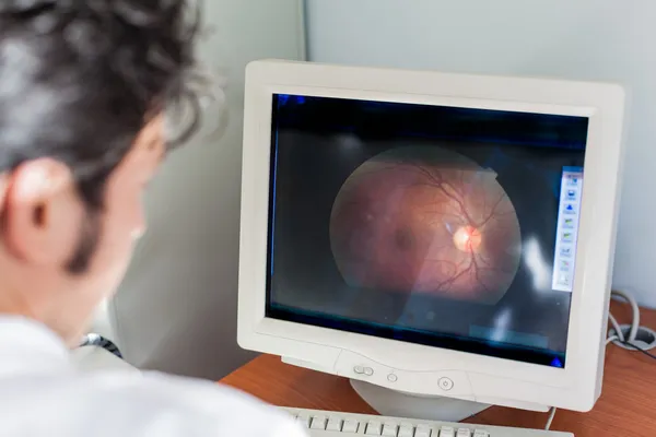
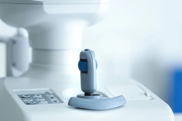
Stock image Fundus Camera
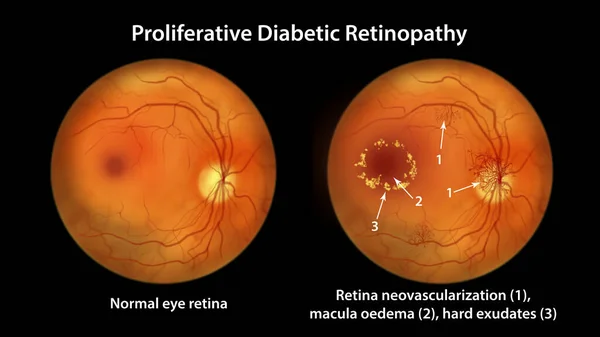
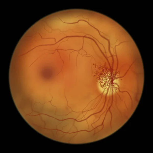
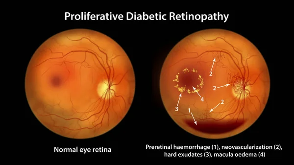
Proliferative Diabetic Retinopathy, Illustration Showing Neovascularization In The Disk And Other Sites, Macula Edema And Hard Exudates. Fundoscopic Examination Of The Eye Retina In Diabetes Mellitus
Image, 6.95MB, 11738 × 6603 jpg


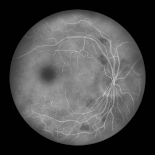
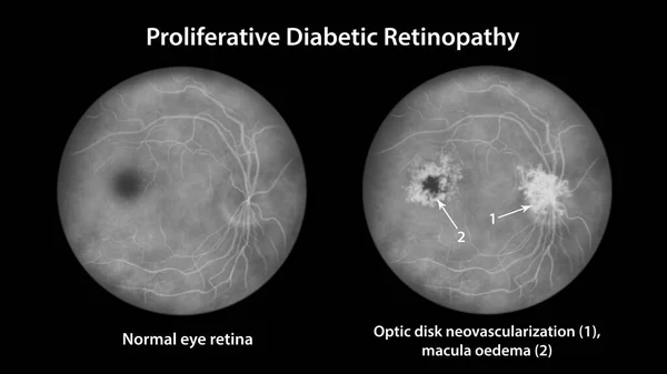
Proliferative Diabetic Retinopathy, Illustration Showing Neovascularization In The Disk And Other Sites, And Macula Edema. Eye Retina In Diabetes Mellitus, Fluorescein Angiography
Image, 2.67MB, 5000 × 5000 jpg


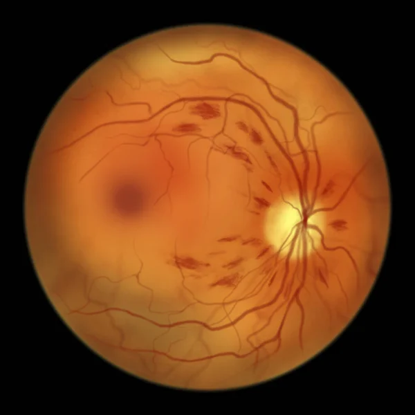
Non-proliferative Diabetic Retinopathy, 3D Illustration Showing Hard Exudates, Microaneurysms, Dot Haemorrhages, Flame-shaped And Splinter Retinal Haemorrhages, Ophthalmoscope View
Image, 13.11MB, 5352 × 5352 jpg

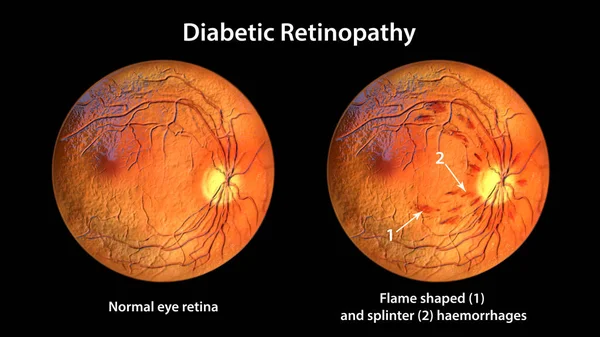
Non-proliferative Diabetic Retinopathy, Illustration Showing Normal Eye Retina And Retina With Hard Exudates, Microaneurysms, Dot Haemorrhages, Flame-shaped And Splinter Retinal Haemorrhages
Image, 7.35MB, 11738 × 6603 jpg

Non-proliferative Diabetic Retinopathy, 3D Illustration Showing Flame-shaped And Splinter Retinal Haemorrhages, Ophthalmoscope View
Image, 13.41MB, 5352 × 5352 jpg

Non-proliferative Diabetic Retinopathy, Illustration Showing Cotton Wool Spots As Fluffy Yellow Patches, Abnormal Finding On Funduscopic Examination Of The Eye Retina In Diabetes Mellitus
Image, 2.7MB, 5000 × 5000 jpg
Non-proliferative Diabetic Retinopathy, Illustration Showing IRMAs (intraretinal Microvascular Abnormalities) As Small Vessels With Abnormal Branching Or Dilatation In Ischaemic Areas
Image, 2.68MB, 5000 × 5000 jpg

Proliferative Diabetic Retinopathy, Illustration Showing Neovascularization In The Optic Disk And Other Sites. Fundoscopic Examination Of The Eye Retina In Diabetes Mellitus, Fluorescein Angiography
Image, 6.07MB, 11738 × 6603 jpg
Non-proliferative Diabetic Retinopathy, Illustration Showing Flame-shaped And Splinter Retinal Haemorrhages, Ophthalmoscope View
Image, 2.96MB, 5000 × 5000 jpg

Non-proliferative Diabetic Retinopathy, Illustration Showing IRMAs (intraretinal Microvascular Abnormalities), Venous Beading, And Microaneurysms
Image, 2.85MB, 5000 × 5000 jpg
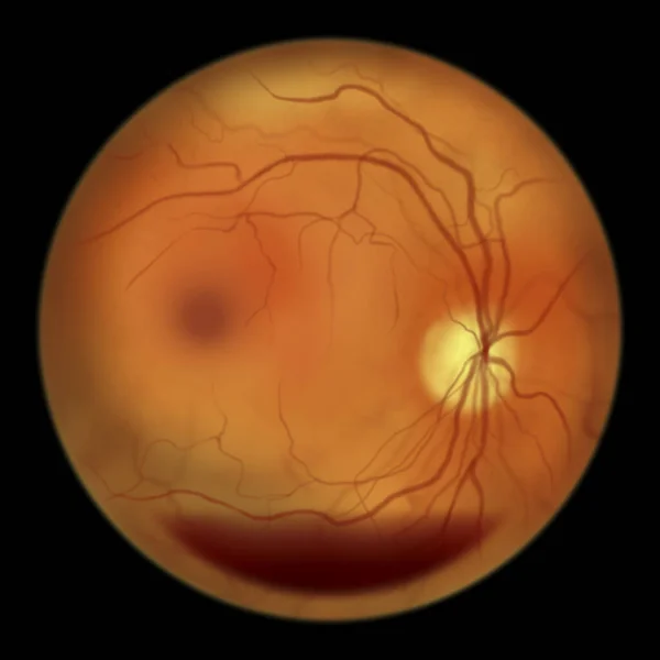
Diabetic Retinopathy, Illustration Shows Preretinal Haemorrhage As Horizontal Blood Level (boat-shaped Haemorrhage), Abnormal Finding On Fundoscopic Examination Of The Eye Retina In Diabetes Mellitus
Image, 2.69MB, 5000 × 5000 jpg

Proliferative Diabetic Retinopathy, Illustration Showing Neovascularization In The Disk And Macula Edema. Abnormal Finding On Fundoscopic Examination Of The Eye Retina In Diabetes Mellitus
Image, 2.85MB, 5000 × 5000 jpg
Proliferative Diabetic Retinopathy, Illustration Showing Neovascularization In The Disk And Other Sites, And Macula Edema. Fundoscopic Examination Of The Eye Retina In Diabetes Mellitus
Image, 2.9MB, 5000 × 5000 jpg

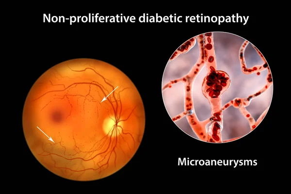
Non-proliferative Diabetic Retinopathy, 3D Illustration Showing Multiple Microaneurysms On The Eye Retina And Closeup View Of Microaneurysms, Microscopic Buldges In The Artery Walls Filled With Blood
Image, 11.11MB, 10431 × 6954 jpg

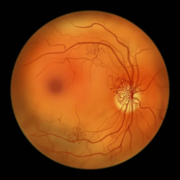
Proliferative Diabetic Retinopathy, Illustration Showing Neovascularization In The Disk And Other Sites, Macula Edema And Hard Exudates. Fundoscopic Examination Of The Eye Retina In Diabetes Mellitus
Image, 3.05MB, 5000 × 5000 jpg
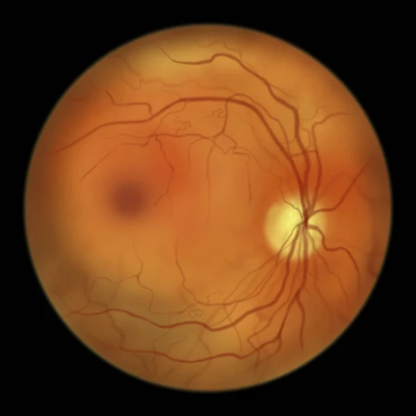
Proliferative Diabetic Retinopathy, Illustration Showing Neovascularization (formation Of New Vessels) In The Optic Disk. Fundoscopic Examination Of The Eye Retina In Diabetes Mellitus
Image, 2.81MB, 5000 × 5000 jpg

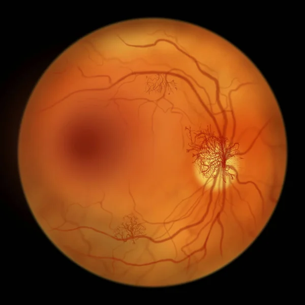
Proliferative Diabetic Retinopathy, Illustration Showing Neovascularization (formation Of New Vessels) In The Optic Disk And Other Sites. Fundoscopic Examination Of The Eye Retina In Diabetes Mellitus
Image, 2.86MB, 5000 × 5000 jpg

Non-proliferative Diabetic Retinopathy, Illustration Showing Normal Eye Retina And Retina With Hard Exudates, Microaneurysms, Dot Haemorrhages, Flame-shaped And Splinter Retinal Haemorrhages
Image, 6.89MB, 11738 × 6603 jpg
Non-proliferative Diabetic Retinopathy, 3D Illustration Showing Multiple Microaneurysms On The Eye Retina And Closeup View Of Microaneurysms, Microscopic Buldges In The Artery Walls Filled With Blood
Image, 10.46MB, 10431 × 6954 jpg
Non-proliferative Diabetic Retinopathy, Illustration Showing Cotton Wool Spots As Fluffy Dark Patches, Abnormal Finding On Funduscopic Examination Of The Eye Retina In Diabetes Mellitus, Fluorescein Angiography
Image, 2.72MB, 5000 × 5000 jpg

Non-proliferative Diabetic Retinopathy, 3D Illustration Showing Normal Eye Retina And Retina With Hard Exudates (irregularly Shaped Yellow Spots)
Image, 25.27MB, 11738 × 6603 jpg

Non-proliferative Diabetic Retinopathy, 3D Illustration Showing Normal Eye Retina And Retina With Hard Exudates, And Cotton Wool Spots
Image, 25.26MB, 11738 × 6603 jpg

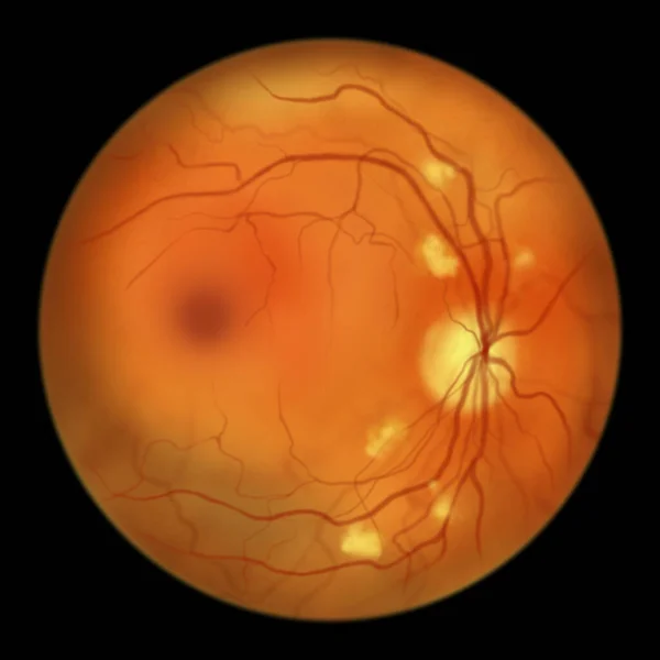
Diabetic Retinopathy, Ophthalmoscope View, Illustration Showing Accumulation Of Fatty Substances Leaked From Blocked Capillaries (yellow Patches), Haemorrhages (red Spots), Microaneurysms
Image, 4.38MB, 5000 × 5000 jpg
Proliferative Diabetic Retinopathy, Illustration Showing Neovascularization In The Disk And Cystoid Macula Edema. Abnormal Finding On Fundoscopic Examination Of The Eye Retina In Diabetes Mellitus
Image, 6.18MB, 11738 × 6603 jpg

Non-proliferative Diabetic Retinopathy, Illustration Showing Normal Eye Retina And Retina With Cotton Wool Spots As Fluffy Yellow Patches
Image, 6.22MB, 11738 × 6603 jpg

Proliferative Diabetic Retinopathy, 3D Illustration Showing Preretinal Haemorrhage As Horizontal Blood Level, Neovascularization In The Disk And Other Sites, Macula Edema And Hard Exudates
Image, 22.98MB, 11738 × 6603 jpg
Proliferative Diabetic Retinopathy, Illustration Showing Preretinal Haemorrhage As Horizontal Blood Level, Neovascularization In The Disk And Other Sites, Macula Edema And Hard Exudates
Image, 7.07MB, 11738 × 6603 jpg
Non-proliferative Diabetic Retinopathy, Ophthalmoscope View, 3D Illustration Showing Normal Eye Retina And Retina With Flame-shaped And Splinter Haemorrhages
Image, 26.2MB, 11738 × 6603 jpg
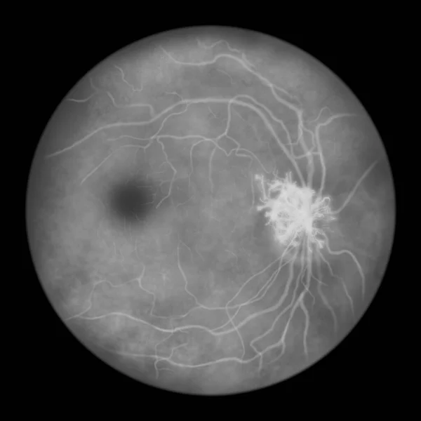
Proliferative Diabetic Retinopathy, Illustration Showing Neovascularization (formation Of New Vessels) In The Optic Disk. Eye Retina In Diabetes Mellitus, Fluorescein Angiography
Image, 2.63MB, 5000 × 5000 jpg


Page 1 >> Next