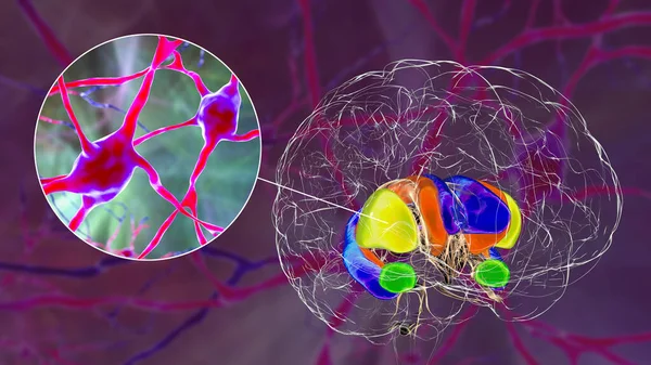
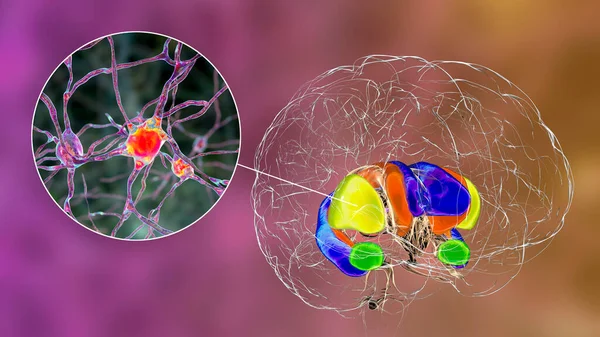
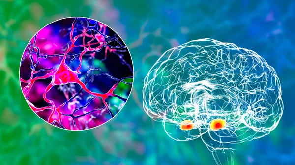
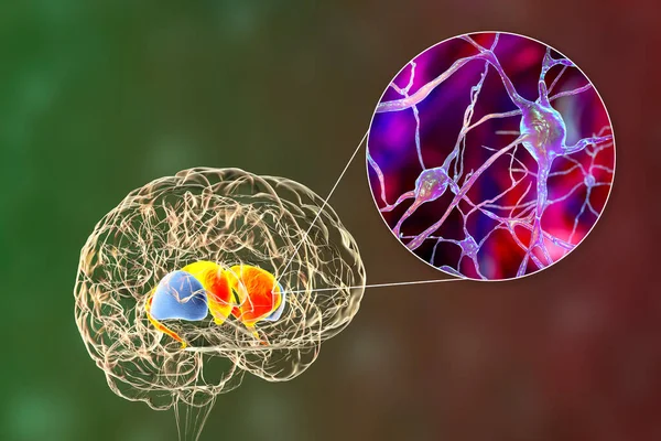
Stock image Dorsal striatum and its neurons in the Huntington's disease, 3D illustration showing enlarged anterior horns of lateral ventricles, degeneration and atrophy of the dorsal striatum, neuronal inclusions

Published: May.24, 2021 13:03:01
Author: katerynakon
Views: 6
Downloads: 0
File type: image / jpg
File size: 17.29 MB
Orginal size: 8515 x 5677 px
Available sizes:
Level: silver