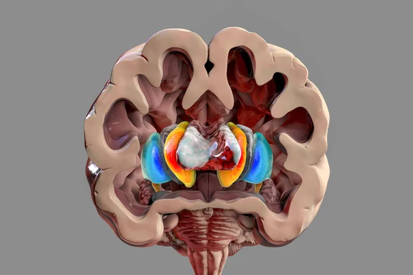
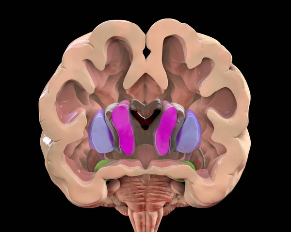
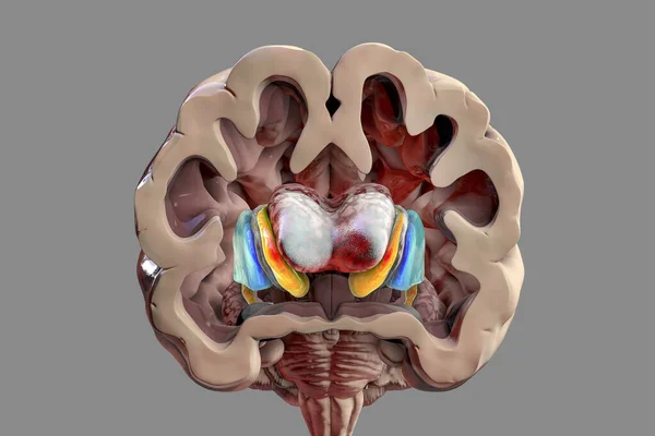
Stock image Dorsal striatum and lateral ventricles in Huntington's disease, 3D illustration showing enlargement of the anterior horns of the lateral ventricles and atrophy of the caudate nuclei
Published: May.24, 2021 13:03:01
Author: katerynakon
Views: 12
Downloads: 0
File type: image / jpg
File size: 7.84 MB
Orginal size: 6342 x 4228 px
Available sizes:
Level: silver