photo free
Home Images Vectors Videos

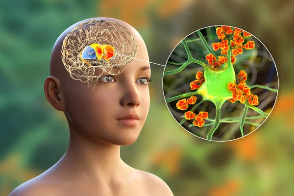
Stock image Dorsal striatum highlighted in child's brain and close-up view of its neurons, 3D illustration. It is a nucleus in the basal ganglia, a component of the motor and reward systems

Stock image Dorsal striatum highlighted in child's brain and close-up view of its neurons, 3D illustration. It is a nucleus in the basal ganglia, a component of the motor and reward systems
Published: May.13, 2021 13:16:57
Author: katerynakon
Views: 18
Downloads: 1
File type: image / jpg
File size: 23.52 MB
Orginal size: 9339 x 6226 px
Available sizes:
S
(500 x 333)
M
(1000 x 667)
L
(2000 x 1333)
XL
(9339 x 6226)
EL0
(9339 x 6226)
Level: silver
Download Image
Image topics:

Similar stock images

stock image Anti-neuronal antibodies, anti-basal ganglia antibodies. 3D illustration shows immunoglobulins attacking neurons in the dorsal striatum of human brain. They are found in post-rheumatic fever chorea

Anti-neuronal Antibodies, Anti-basal Ganglia Antibodies. 3D Illustration Shows Immunoglobulins Attacking Neurons In The Dorsal Striatum Of Human Brain. They Are Found In Post-rheumatic Fever Chorea

9339 × 6226
stock image Dorsal striatum highlighted in child's brain and close-up view of its neurons, 3D illustration. It is a nucleus in the basal ganglia, a component of the motor and reward systems

Dorsal Striatum Highlighted In Child's Brain And Close-up View Of Its Neurons, 3D Illustration. It Is A Nucleus In The Basal Ganglia, A Component Of The Motor And Reward Systems

9339 × 6226
stock image Brain dorsal striatum anatomy, 3D illustration. The dorsal striatum consists of the caudate nucleus (green) and the putamen (orange). Amygdala is colored in blue

Brain Dorsal Striatum Anatomy, 3D Illustration. The Dorsal Striatum Consists Of The Caudate Nucleus (green) And The Putamen (orange). Amygdala Is Colored In Blue

6000 × 4000
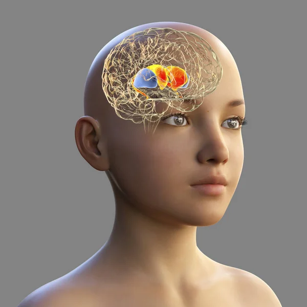
stock image Dorsal striatum in the child's brain, 3D illustration. It is a nucleus in the basal ganglia, consists of the caudate nucleus (red) and the putamen (blue), is a component of the motor and reward system

Dorsal Striatum In The Child's Brain, 3D Illustration. It Is A Nucleus In The Basal Ganglia, Consists Of The Caudate Nucleus (red) And The Putamen (blue), Is A Component Of The Motor And Reward System

6109 × 6109
stock image Amygdala in the brain, and closeup view of amygdala neurons, 3D illustration. Two almond-shaped clusters of nuclei within temporal lobes, part of the limbic system, play role in memory and emotions

Amygdala In The Brain, And Closeup View Of Amygdala Neurons, 3D Illustration. Two Almond-shaped Clusters Of Nuclei Within Temporal Lobes, Part Of The Limbic System, Play Role In Memory And Emotions

6000 × 4000
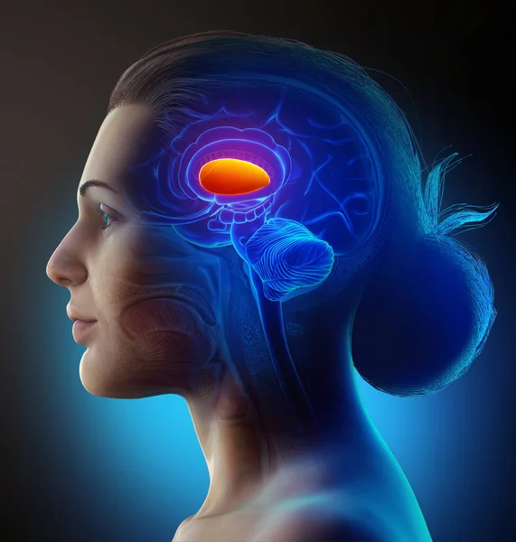
stock image 3d rendered medically accurate illustration of a female  brains  anatomy- the putamen

3d Rendered Medically Accurate Illustration Of A Female Brains Anatomy- The Putamen

4000 × 4200
stock image Anti-basal ganglia antibodies. 3D conceptual illustration showing molecules of immunoglobulins attacking dorsal striatum highlighted in the child's brain. They are found in post-rheumatic fever chorea

Anti-basal Ganglia Antibodies. 3D Conceptual Illustration Showing Molecules Of Immunoglobulins Attacking Dorsal Striatum Highlighted In The Child's Brain. They Are Found In Post-rheumatic Fever Chorea

9828 × 6552