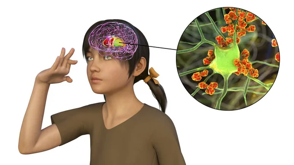
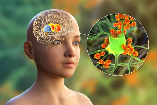
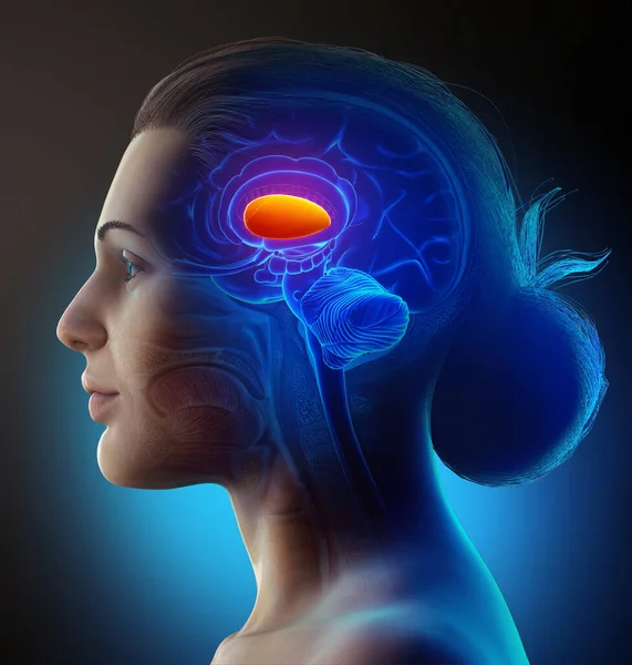
Stock image Dorsal striatum highlighted in child's brain and close-up view of its neurons, 3D illustration. It is a nucleus in the basal ganglia, a component of the motor and reward systems

Published: May.13, 2021 13:16:57
Author: katerynakon
Views: 101
Downloads: 4
File type: image / jpg
File size: 31.18 MB
Orginal size: 9339 x 6226 px
Available sizes:
Level: silver