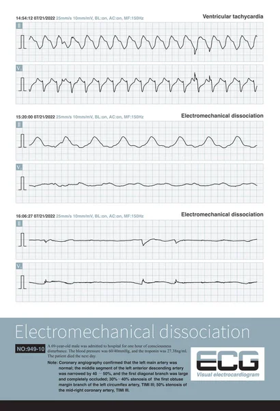
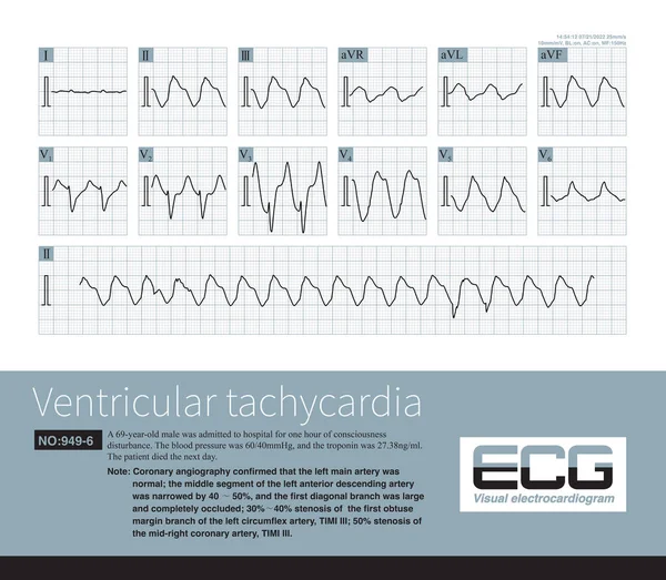
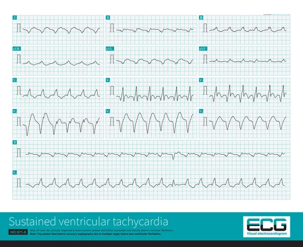
Stock image Electromechanical separation is a kind of terminal ECG. The patient's ECG has electrical signals, the ECG wave is widened with morphological abnormalities, and the ventricle has no contraction.

Published: Apr.11, 2023 09:26:03
Author: asia11m
Views: 95
Downloads: 1
File type: image / jpg
File size: 21.68 MB
Orginal size: 10000 x 10515 px
Available sizes:
Level: beginner