photo free
Home Images Vectors Videos

Stock image Ventricular Tachycardia

stock vector Schemes set of common electrocardiogram (ECG) abnormalities, including partial blocks and flutter

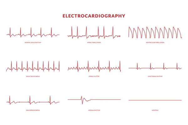
Schemes Set Of Common Electrocardiogram (ECG) Abnormalities, Including Partial Blocks And Flutter

Vector, 9.68MB, 7750 × 4367 eps
stock vector Electrocardiography Heartbeat Line monitor. vector EPS10 Illustration

Electrocardiography Heartbeat Line Monitor. Vector EPS10 Illustration

Vector, 0.82MB, 6000 × 4000 eps
stock image Ventricular Tachycardia -  is fast heart rhythm, that originates in one of the ventricles of the heart. This is a potentially life-threatening arrhythmia because it may lead to ventricular fibrillation, asystole, and sudden death

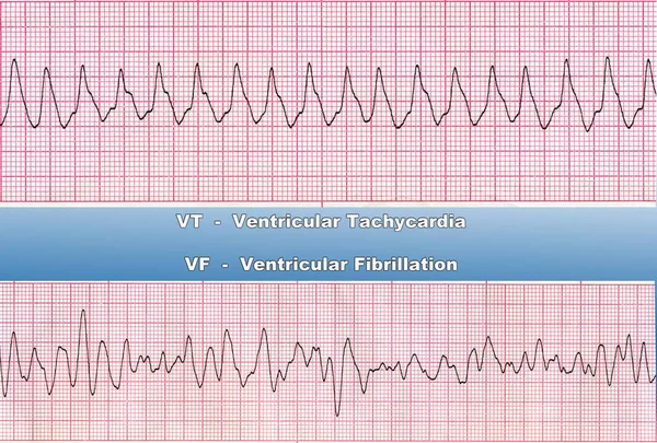
Ventricular Tachycardia - Is Fast Heart Rhythm, That Originates In One Of The Ventricles Of The Heart. This Is A Potentially Life-threatening Arrhythmia Because It May Lead To Ventricular Fibrillation, Asystole, And Sudden Death

Image, 7.23MB, 3935 × 2663 jpg
stock image Ventricular Tachycardia - Deadly Heart Rhythm

Ventricular Tachycardia - Deadly Heart Rhythm

Image, 5.4MB, 3935 × 2331 jpg
stock vector Heart structure. Types of cardiac arrhythmia.

Heart Structure. Types Of Cardiac Arrhythmia.

Vector, 8.45MB, 5306 × 5292 eps
stock image Defibrillator practice on a CPR

Defibrillator Practice On A CPR

Image, 7.67MB, 5184 × 3456 jpg
stock image Electric plug with money and coins.

Electric Plug With Money And Coins.

Image, 9.03MB, 4600 × 3067 jpg
stock image Male, 84 years old, admitted to hospital with chest pain for 1 day. ECG showed acute inferior and posterior MI and possibly right MI. The patient died of ventricular fibrillation the next day.

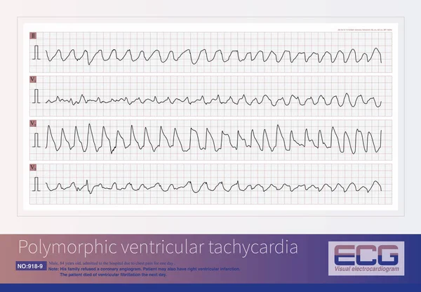
Male, 84 Years Old, Admitted To Hospital With Chest Pain For 1 Day. ECG Showed Acute Inferior And Posterior MI And Possibly Right MI. The Patient Died Of Ventricular Fibrillation The Next Day.

Image, 17.76MB, 10000 × 6934 jpg
stock image Ventricular tachyarrhythmia includes many clinical types, some benign and some malignant. For malignant ventricular arrhythmias, patients are at risk of death.

Ventricular Tachyarrhythmia Includes Many Clinical Types, Some Benign And Some Malignant. For Malignant Ventricular Arrhythmias, Patients Are At Risk Of Death.

Image, 27.66MB, 8000 × 10973 jpg
stock image Electromechanical separation is a kind of terminal ECG. The patient's ECG has electrical signals, the ECG wave is widened with morphological abnormalities, and the ventricle has no contraction.

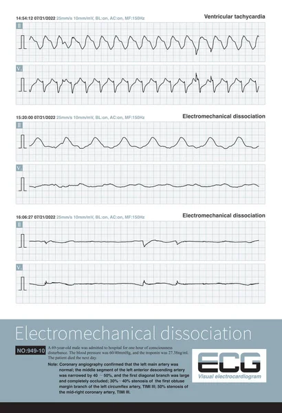
Electromechanical Separation Is A Kind Of Terminal ECG. The Patient's ECG Has Electrical Signals, The ECG Wave Is Widened With Morphological Abnormalities, And The Ventricle Has No Contraction.

Image, 21.68MB, 10000 × 10515 jpg
stock image Torsade de pointes refers to the pleomorphic ventricular tachycardia that occurs in the background of long QT interval, and the polarity of QRS wave twists around the equipotential line.

Torsade De Pointes Refers To The Pleomorphic Ventricular Tachycardia That Occurs In The Background Of Long QT Interval, And The Polarity Of QRS Wave Twists Around The Equipotential Line.

Image, 18.28MB, 10000 × 5808 jpg
stock image A patient with acute extensive anterior  myocardial infarction developed ventricular tachycardia during hospitalization and quickly experienced cardiac arrest.

A Patient With Acute Extensive Anterior Myocardial Infarction Developed Ventricular Tachycardia During Hospitalization And Quickly Experienced Cardiac Arrest.

Image, 31.98MB, 10000 × 14632 jpg
stock image Electrocardiograph Traces - Cardiac Arrhythmia

Electrocardiograph Traces - Cardiac Arrhythmia

Image, 11.43MB, 3413 × 5120 jpg
stock image The illustration shows the two patterns of ventricular tachycardia episodes.The green circle represents sinus rhythm. Picture A shows paroxysmal episodes of ventricular tachycardia, and picture B shows short bursts.

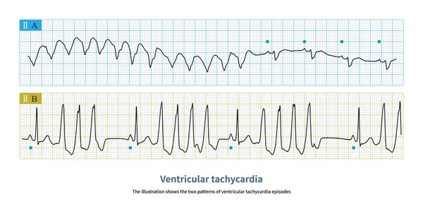
The Illustration Shows The Two Patterns Of Ventricular Tachycardia Episodes.The Green Circle Represents Sinus Rhythm. Picture A Shows Paroxysmal Episodes Of Ventricular Tachycardia, And Picture B Shows Short Bursts.

Image, 10.72MB, 10000 × 5059 jpg
stock vector Amiodarone molecule. It is antiarrhythmic, vasodilatory, cardiovascular drug. Structural chemical formula and molecule model. Sheet of paper in a cage. Vector illustration

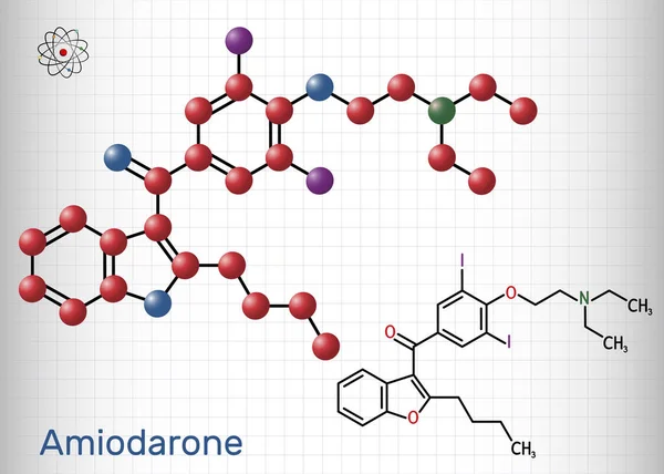
Amiodarone Molecule. It Is Antiarrhythmic, Vasodilatory, Cardiovascular Drug. Structural Chemical Formula And Molecule Model. Sheet Of Paper In A Cage. Vector Illustration

Vector, 5.45MB, 5916 × 4226 eps
stock image A patient with AIMI presents with a sudden widening of the QRS complex in the junctional escape rhythm, premature ventricular contractions, resulting in  polymorphic ventricular tachycardia.

A Patient With AIMI Presents With A Sudden Widening Of The QRS Complex In The Junctional Escape Rhythm, Premature Ventricular Contractions, Resulting In Polymorphic Ventricular Tachycardia.

Image, 14.66MB, 10000 × 7554 jpg
stock image ECG tape with paroxysmal ventricular tachycardia and ventricular asystole

ECG Tape With Paroxysmal Ventricular Tachycardia And Ventricular Asystole

Image, 5.17MB, 2800 × 1915 jpg
stock image ECG tape with paroxysmal ventricular tachycardia

ECG Tape With Paroxysmal Ventricular Tachycardia

Image, 7.6MB, 3189 × 2000 jpg
stock image Businessman using laptop computer overnight cause heart attack failure symptom. Healthcare and medical wellness of overworked people lifestyle concept. Technology and workaholic illness theme.

Businessman Using Laptop Computer Overnight Cause Heart Attack Failure Symptom. Healthcare And Medical Wellness Of Overworked People Lifestyle Concept. Technology And Workaholic Illness Theme.

Image, 14.75MB, 6016 × 4016 jpg
stock vector Vector medical illustration with emblem of tachycardia

Vector Medical Illustration With Emblem Of Tachycardia

Vector, 2.17MB, 4167 × 4167 eps
stock vector Heart Defibrillator flat icon with long shadow

Heart Defibrillator Flat Icon With Long Shadow

Vector, 1.07MB, 6005 × 6005 eps
stock vector Heart Defibrillator flat icon with long shadow

Heart Defibrillator Flat Icon With Long Shadow

Vector, 4.58MB, 6005 × 6005 eps
stock image Treatment, support with medication and heart protection. Drugs - vials and syringe on red background aimed at heart, which lies nearby. For use in cardiology and treatment of cardiovascular system

Treatment, Support With Medication And Heart Protection. Drugs - Vials And Syringe On Red Background Aimed At Heart, Which Lies Nearby. For Use In Cardiology And Treatment Of Cardiovascular System

Image, 4.65MB, 6016 × 4000 jpg
stock image Diagnosis, treatment and prevention of diseases of heart and cardiovascular system concept photo. Blue stethoscope is surrounded by tape of ECG with electrocardiogram drawn on it. Diagnosis of disease

Diagnosis, Treatment And Prevention Of Diseases Of Heart And Cardiovascular System Concept Photo. Blue Stethoscope Is Surrounded By Tape Of ECG With Electrocardiogram Drawn On It. Diagnosis Of Disease

Image, 6.13MB, 6000 × 4000 jpg
stock vector AED - automated external defibrillator    device

AED - Automated External Defibrillator Device

Vector, 7.15MB, 6320 × 6320 eps
stock vector Heart Defibrillator flat icon with long shadow

Heart Defibrillator Flat Icon With Long Shadow

Vector, 4.6MB, 6005 × 6005 eps
stock vector Healthy heart with arrhythmogenic cardiomyopathy. Vector illustration of arrhytmogenic right ventricular dysplasia

Healthy Heart With Arrhythmogenic Cardiomyopathy. Vector Illustration Of Arrhytmogenic Right Ventricular Dysplasia

Vector, 7.18MB, 4910 × 4000 eps
stock image When the left free wall and septal accessory pathway are excited, preexcitation waves with different polarities are generated in leads  and aVL.

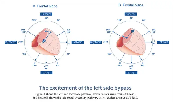
When The Left Free Wall And Septal Accessory Pathway Are Excited, Preexcitation Waves With Different Polarities Are Generated In Leads And AVL.

Image, 6.98MB, 14851 × 8810 jpg
stock vector Heart defibrillator

Heart Defibrillator

Vector, 0.28MB, 2800 × 2800 eps
stock vector Heart Defibrillator flat icon with long shadow

Heart Defibrillator Flat Icon With Long Shadow

Vector, 4.58MB, 6005 × 6005 eps
stock vector Heart Defibrillator flat icon with long shadow

Heart Defibrillator Flat Icon With Long Shadow

Vector, 1.03MB, 6005 × 6005 eps
stock image Ventricular Fibrillation - Deadly Heart Rhythm

Ventricular Fibrillation - Deadly Heart Rhythm

Image, 5.27MB, 3935 × 2337 jpg
stock image Automated external defibrillator, automatic electronic defibrillator AED, 3D rendering isolated on white background

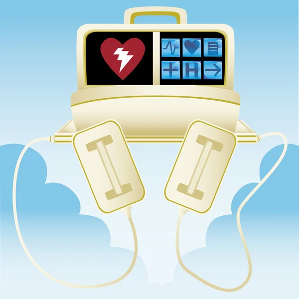
Automated External Defibrillator, Automatic Electronic Defibrillator AED, 3D Rendering Isolated On White Background

Image, 5.19MB, 10000 × 6500 jpg
stock image A 4-year-old boy with a clinical diagnosis of long QT syndrome. No genetic testing was done during hospitalization. The child died suddenly during follow-up.

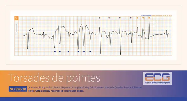
A 4-year-old Boy With A Clinical Diagnosis Of Long QT Syndrome. No Genetic Testing Was Done During Hospitalization. The Child Died Suddenly During Follow-up.

Image, 7.66MB, 10000 × 5414 jpg
stock vector Sotalol

Sotalol

Vector, 0.93MB, 6320 × 4000 eps
stock image Chest compressions on a simulation dummy during basic life support with an automatic external defibrillator. Simulation training scenario. Vertical

Chest Compressions On A Simulation Dummy During Basic Life Support With An Automatic External Defibrillator. Simulation Training Scenario. Vertical

Image, 3.99MB, 2567 × 3850 jpg
stock image ECG paper graph report, electrocardiogram on paper form as background

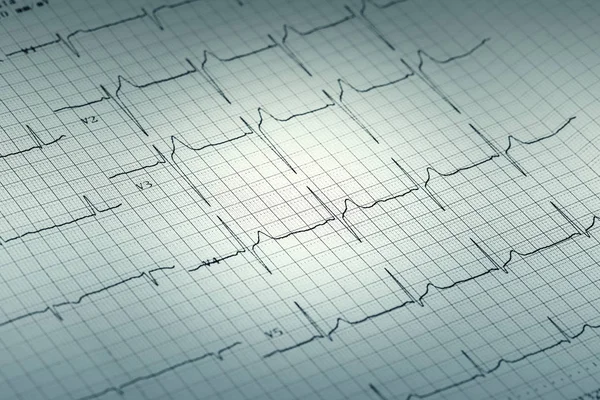
ECG Paper Graph Report, Electrocardiogram On Paper Form As Background

Image, 10.21MB, 6000 × 4000 jpg
stock image Automated external defibrillator, 3D rendering isolated on white background

Automated External Defibrillator, 3D Rendering Isolated On White Background

Image, 5.15MB, 8000 × 5300 jpg
stock image Defibrillator

Defibrillator

Image, 3.65MB, 4016 × 2677 jpg
stock image On the electrocardiogram, observing the morphology of QRS waves in lead V1 can distinguish whether ventricular pre excitation is located in the left ventricle or the right ventricle.

On The Electrocardiogram, Observing The Morphology Of QRS Waves In Lead V1 Can Distinguish Whether Ventricular Pre Excitation Is Located In The Left Ventricle Or The Right Ventricle.

Image, 4.78MB, 10000 × 11226 jpg
stock image Automated external defibrillator, AED. 3D rendering isolated on white background

Automated External Defibrillator, AED. 3D Rendering Isolated On White Background

Image, 6.64MB, 10000 × 6500 jpg
stock image a pacemaker lies directly next to a defibrillator on a green surgical drape

A Pacemaker Lies Directly Next To A Defibrillator On A Green Surgical Drape

Image, 27.64MB, 6000 × 4000 jpg
stock vector Heart Defibrillator flat icon with long shadow

Heart Defibrillator Flat Icon With Long Shadow

Vector, 4.59MB, 6005 × 6005 eps
stock vector Defibrillator in flat. Device for restoring the heartbeat

Defibrillator In Flat. Device For Restoring The Heartbeat

Vector, 0.78MB, 4000 × 4000 eps
stock image Hand with pen drawing the chemical formula of Amiodarone

Hand With Pen Drawing The Chemical Formula Of Amiodarone

Image, 5.14MB, 6000 × 4000 jpg
stock vector Heart Defibrillator flat icon with long shadow

Heart Defibrillator Flat Icon With Long Shadow

Vector, 1.01MB, 6005 × 6005 eps
stock image When the ventricular preexcitation wave leaves the baseline and then falls back to the baseline, it is interpreted as an isoelectric line preexcitation wave.

When The Ventricular Preexcitation Wave Leaves The Baseline And Then Falls Back To The Baseline, It Is Interpreted As An Isoelectric Line Preexcitation Wave.

Image, 9.3MB, 10000 × 11275 jpg
stock image icd word written on wood block. international classification diseases text on table, concept.

Icd Word Written On Wood Block. International Classification Diseases Text On Table, Concept.

Image, 6.13MB, 3800 × 2534 jpg
stock image When the left anterior wall and posterior wall accessory pathway are excited, preexcitation waves with different polarities are generated in the inferior wall leads of ,  and aVF.

When The Left Anterior Wall And Posterior Wall Accessory Pathway Are Excited, Preexcitation Waves With Different Polarities Are Generated In The Inferior Wall Leads Of , And AVF.

Image, 3.66MB, 10000 × 5932 jpg
stock image Defibrillator

Defibrillator

Image, 2.38MB, 2007 × 3010 jpg
Page 1 >> Next