Home
Images
Vectors
Videos
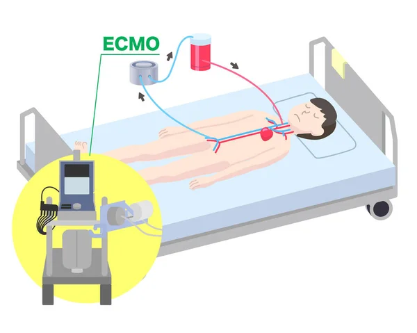
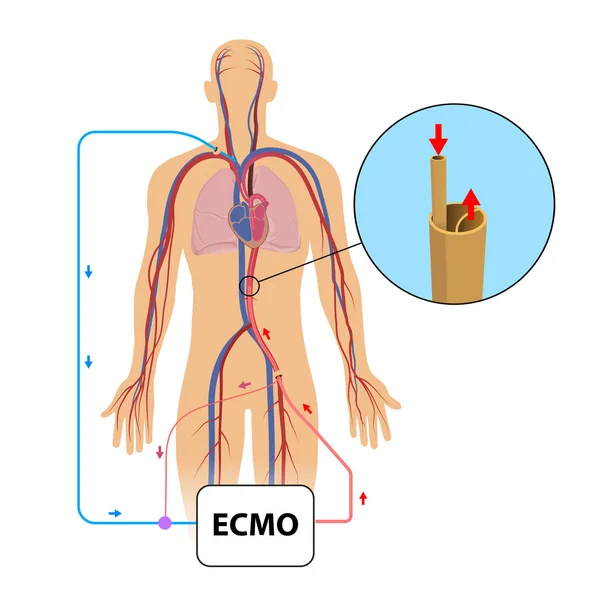
Stock image Extracorporeal membrane oxygenation,ecmo in intensive care department
Published:
Mar.03, 2020 09:50:15
Author:
madrock24
Views:
89
Downloads:
5
File type:
image
/
jpg
File size:
1.87 MB
Orginal size:
5000
x
5000
px
Available sizes:
S
(500 x 500)
M
(1000 x 1000)
L
(2000 x 2000)
XL
(5000 x 5000)
EL0
(5000 x 5000)
Level:
beginner
Download Image
Image topics:
White
Illustration
Equipment
Blood
Medicine
Healthcare
Medical
Care
Line
Machine
Industry
Emergency
Heart
Hospital
Patient
Room
Disease
Vessel
Membrane
Anatomy
Surgery
Plaque
Monitoring
Operation
Cardio
Bypass
Extra
Cardiac
Intensive
Vascular
Infarction
Intervention
Aorta
Myocardial
ICU
Coronary
Cannula
Catheter
Atherosclerosis
Health Care
Heart Attack
Oxygenation
Extracorporeal
ECMO
Coronary Arteries
Oxygenator
Stemi
Ecmo Machine
Medical Ecmo
Similar stock images
Extracorporeal Membrane Oxygenator Illustration
5000 × 4000
ECMO ( Extracorporeal Membrane Oxygenation ) Structure Vector Illustration
7684 × 6537
Atrial Depolarization Forms A P Wave, Ventricular Depolarization Forms A QRS Wave, And Ventricular Repolarization Forms A T Wave, Which Constitutes A Common Waveform Of The Electrocardiogram.
10000 × 9917
Extracorporeal Membrane Oxygenation. Diagram For Explain Rescue Patients Respiratory Failure By Using Instrumental Extracorporeal Membrane Oxygenation ECMO
5000 × 5000
Medical Illustration Of The Two Main Phases Of The Cardiac Cycle, Systole And Diastole, With Annotations.
3427 × 3308
Medical Illustration Of ECG & The Electrical Activity Of The Heart
8031 × 8410
Tension Pneumothorax Disease. Reducing The Amount Of Blood Returned To The Heart. Lung Or Chest Wall Injury. Chest Pain, Shortness Of Breathing. Unhealthy Internal Organs In Respiratory System Vector
4168 × 4168
The Positive Electrode Of Lead Of The ECG Is Placed On The Left Lower Extremity, And The Negative Electrode Is Placed On The Left Upper Extremity, Which Is A Standard Bipolar Limb Lead.
9000 × 11537