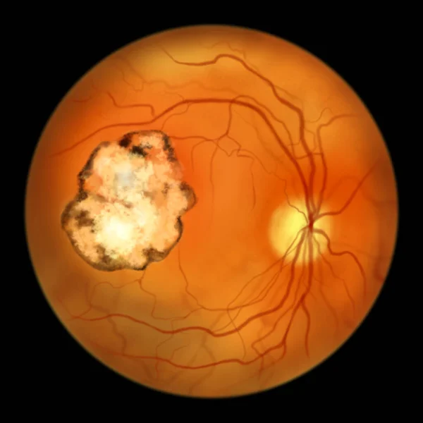
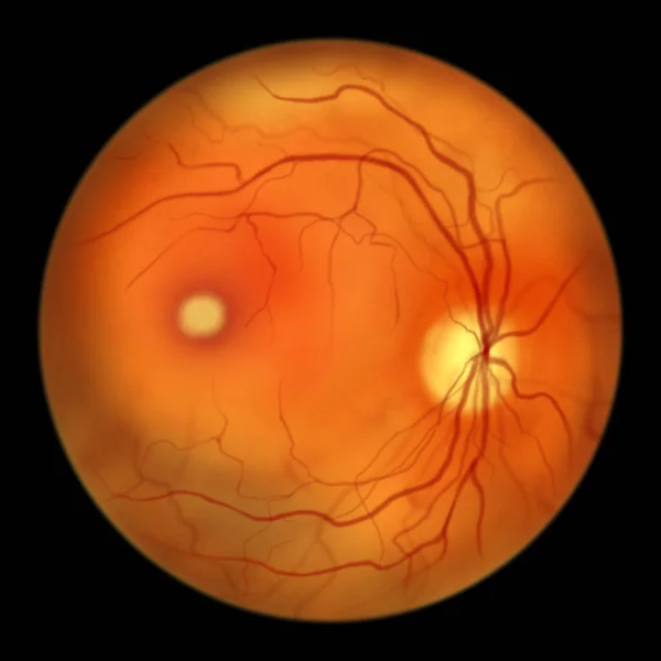
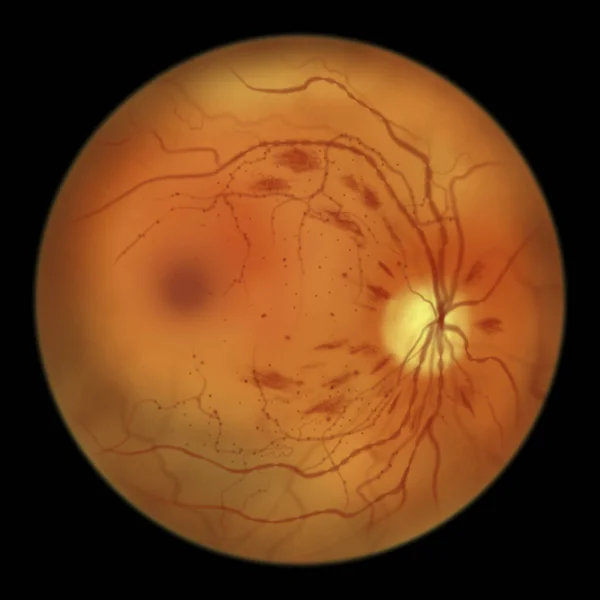
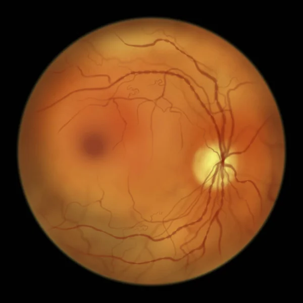
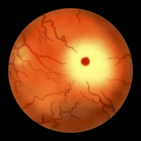
Stock image Eye retina in Tay-Sachs disease, 3D illustration with so-called cherry-red spot. A genetic disorder that progressively destroys brain neurons, is caused by a genetic mutation in the HEXA gene
Published: Oct.12, 2021 09:05:24
Author: katerynakon
Views: 20
Downloads: 1
File type: image / jpg
File size: 3.82 MB
Orginal size: 5000 x 5000 px
Available sizes:
Level: silver