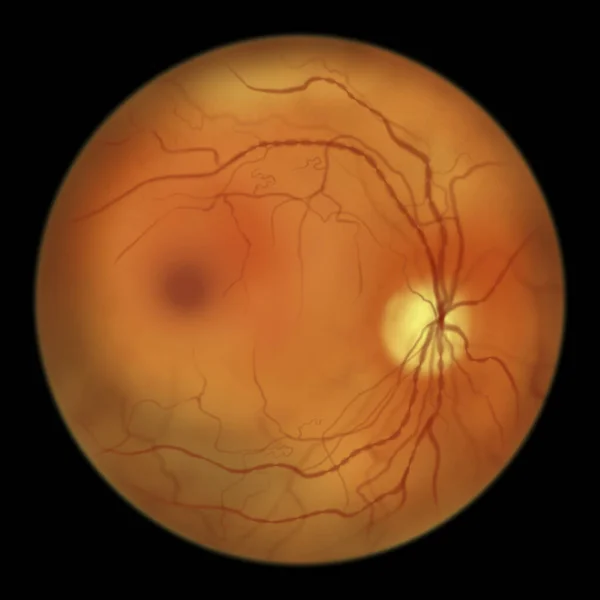
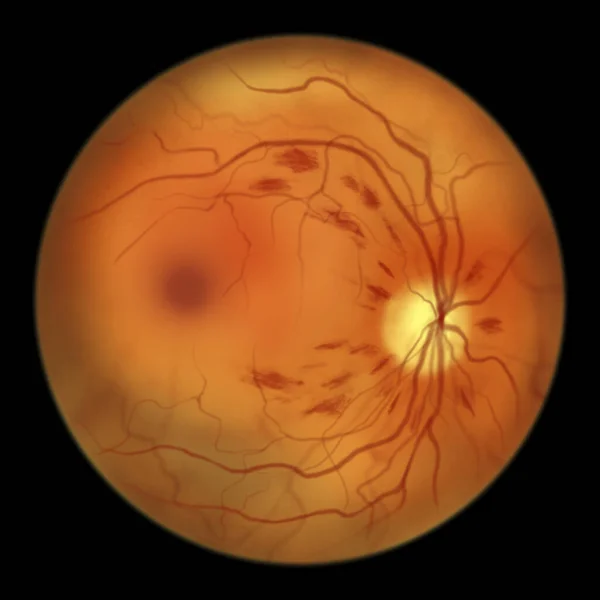
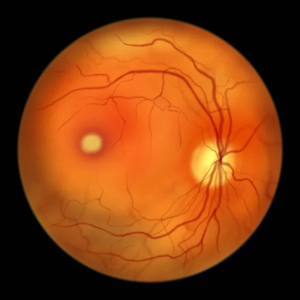
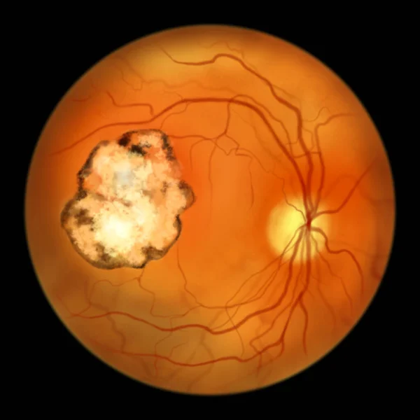
Stock image Retinal scar in toxoplasmosis, a disease caused by the single-celled protozoan Toxoplasma gondii, ophthalmoscope view, scientific illustration
Published: Mar.20, 2023 10:06:05
Author: katerynakon
Views: 19
Downloads: 1
File type: image / jpg
File size: 3.02 MB
Orginal size: 5000 x 5000 px
Available sizes:
Level: silver