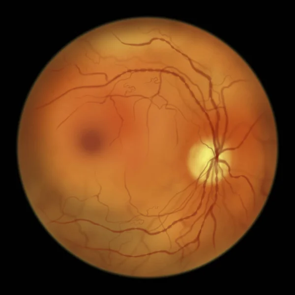
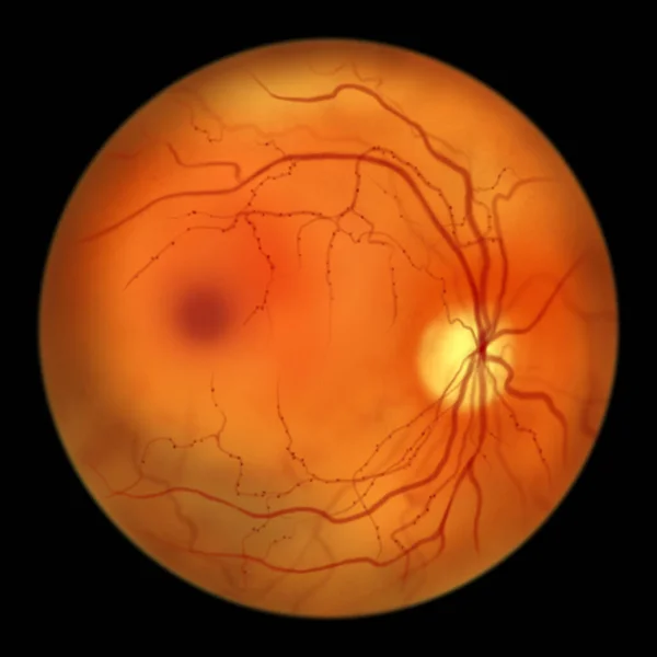
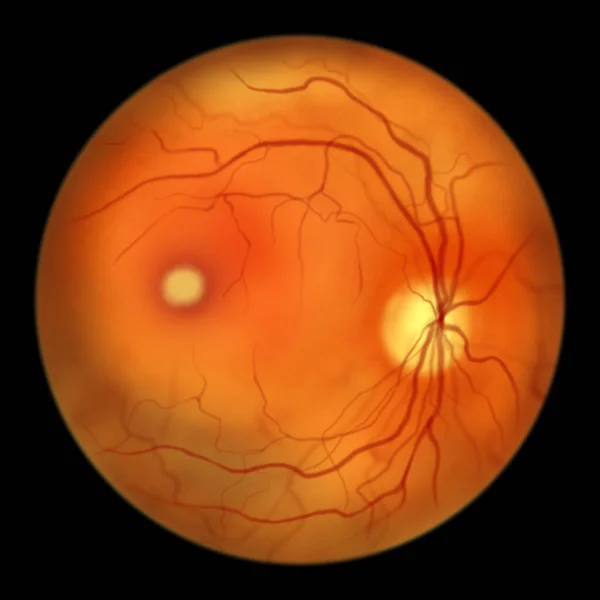
Stock image Best disease. Best vitelliform macular dystrophy, Vitelliform stage, classic egg-yolk lesion, scientific illustration, ophthalmoscope view
Published: Mar.16, 2023 08:26:23
Author: katerynakon
Views: 23
Downloads: 1
File type: image / jpg
File size: 2.87 MB
Orginal size: 5000 x 5000 px
Available sizes:
Level: silver
Similar stock images

Non-proliferative Diabetic Retinopathy, Illustration Showing Venous Beading, Ophthalmoscope View
5000 × 5000