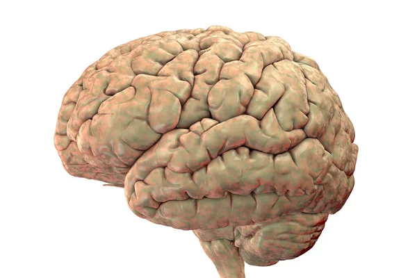
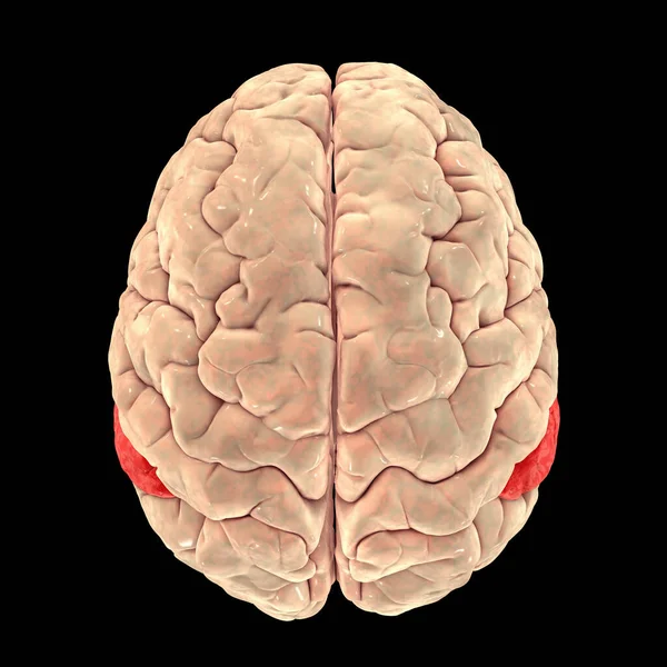
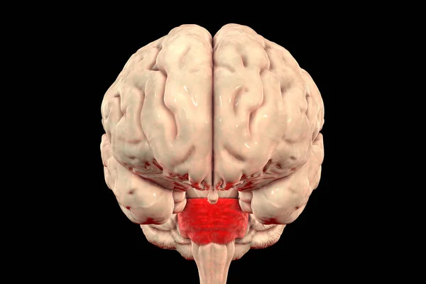
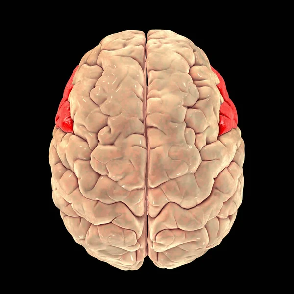
Stock image Human brain anatomical model isolated on white background, realistic 3d illustration

Published: Jun.18, 2021 08:44:25
Author: katerynakon
Views: 22
Downloads: 1
File type: image / jpg
File size: 4.04 MB
Orginal size: 6000 x 4000 px
Available sizes:
Level: silver