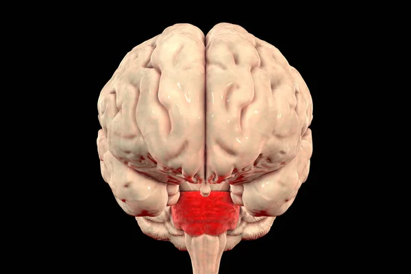
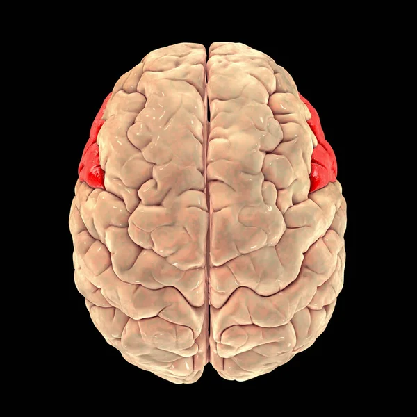
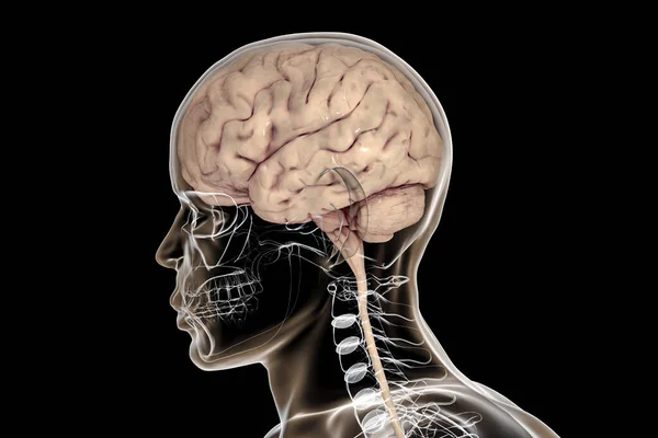
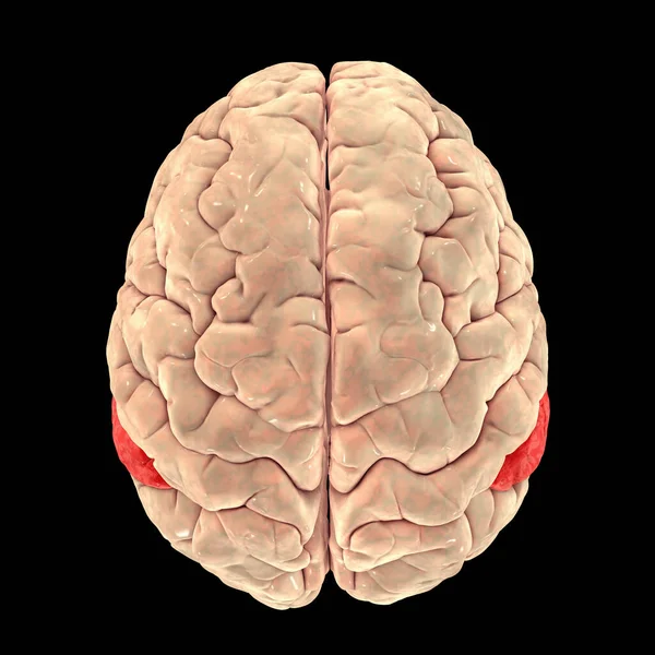
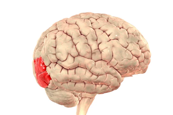
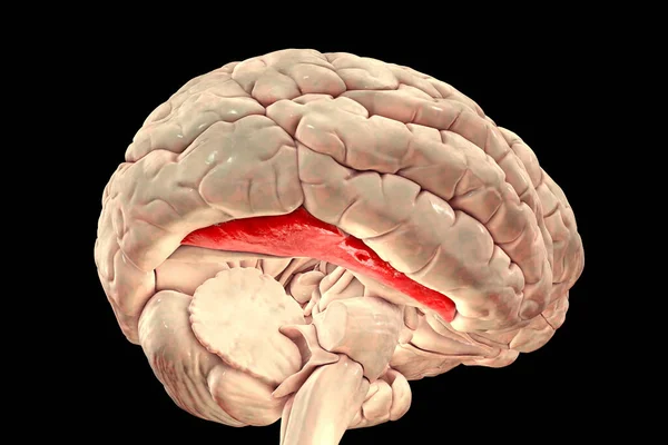
Stock image Human brain with highlighted fusiform gyrus, or medial occipitotemporal gyrus, 3D illustration. It is associated with various neural pathways related to recognition, and also to synesthesia, dyslexia, and prosopagnosia
Published: Nov.13, 2020 11:56:38
Author: katerynakon
Views: 14
Downloads: 1
File type: image / jpg
File size: 4.67 MB
Orginal size: 6000 x 4000 px
Available sizes:
Level: silver