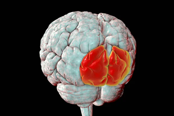
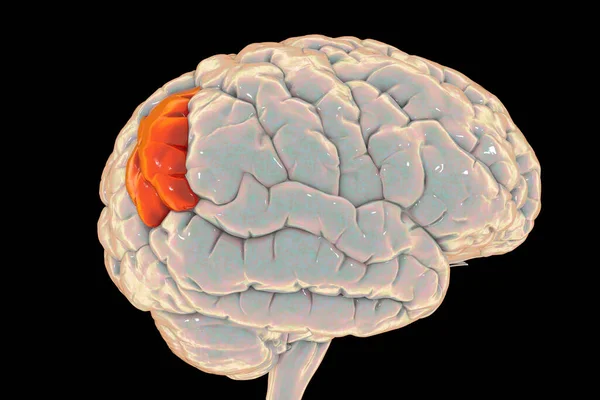
Stock image Human brain with highlighted lingual gyrus, or medial occipitotemporal gyrus, inferior view, 3D illustration. It plays an important role in vision and dreaming

Published: Nov.13, 2020 11:56:38
Author: katerynakon
Views: 15
Downloads: 0
File type: image / jpg
File size: 5.31 MB
Orginal size: 5000 x 5000 px
Available sizes:
Level: silver