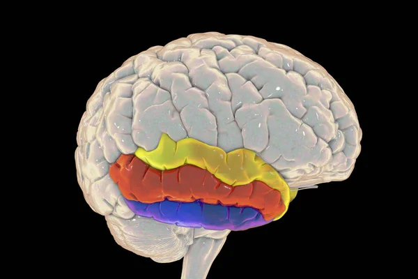
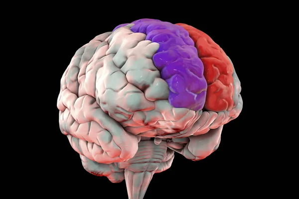
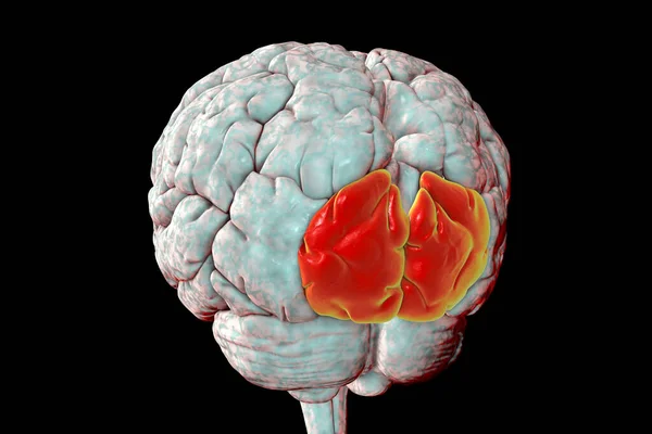
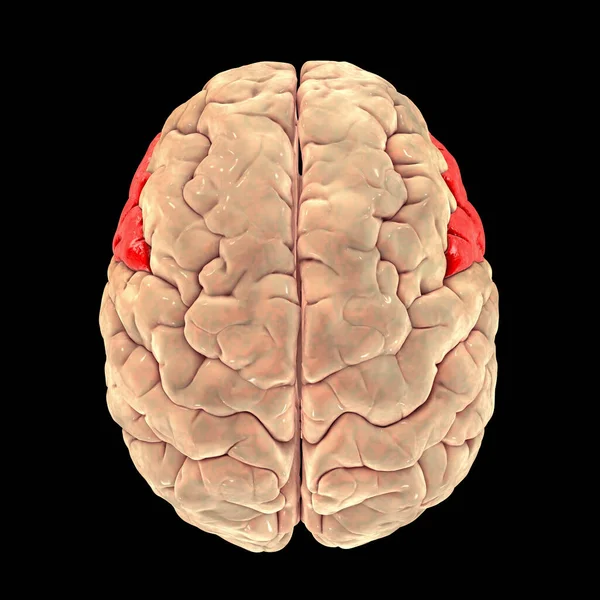
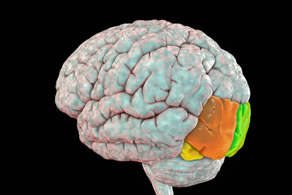
Stock image Human brain with highlighted occipital gyri superior (green), middle (orange), and inferior (yellow), 3D illustration. Also known as the occipital face area, they are responsible for object recognition
Published: Dec.01, 2020 16:21:16
Author: katerynakon
Views: 12
Downloads: 0
File type: image / jpg
File size: 5.85 MB
Orginal size: 6000 x 4000 px
Available sizes:
Level: silver