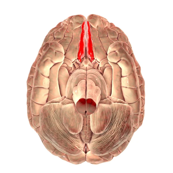
Stock image Human brain with highlighted straight gyrus, or gyrus rectus, inferior view, 3D illustration. It is located in the inferior frontal lobe
Published: Dec.01, 2020 16:21:16
Author: katerynakon
Views: 15
Downloads: 1
File type: image / jpg
File size: 7.77 MB
Orginal size: 6000 x 6000 px
Available sizes:
Level: silver