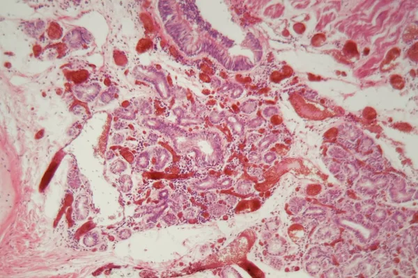
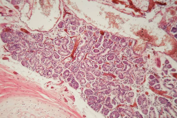
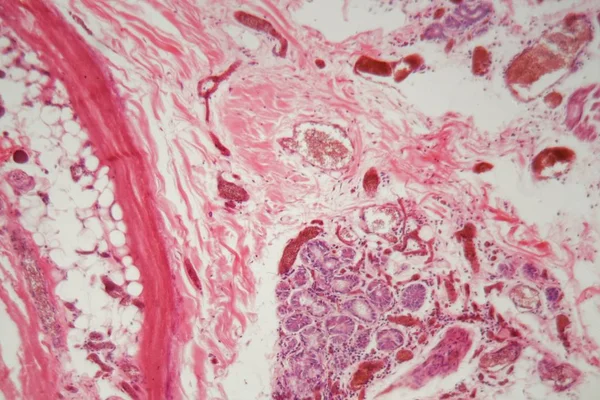
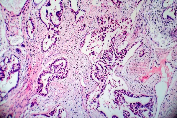
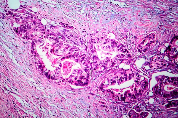
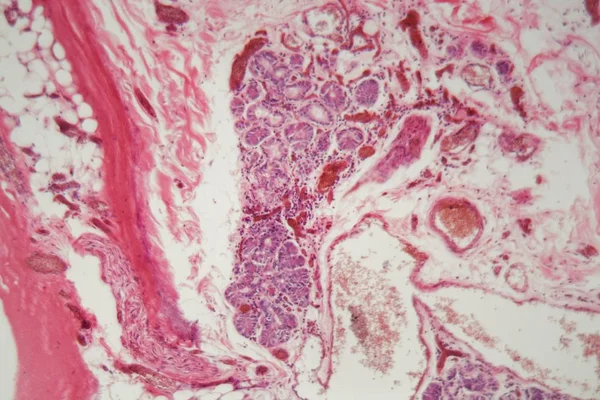
Stock image Human lung tissue with Pulmonary embolism under a microscope.
Published: Nov.27, 2019 09:14:42
Author: ChWeiss
Views: 12
Downloads: 0
File type: image / jpg
File size: 1.66 MB
Orginal size: 5616 x 3744 px
Available sizes:
Level: bronze