Home
Images
Vectors
Videos
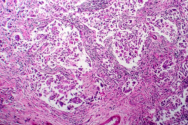
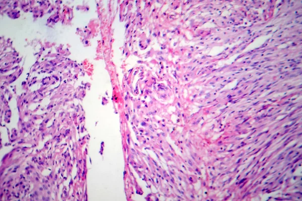
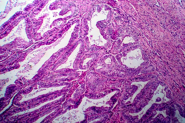
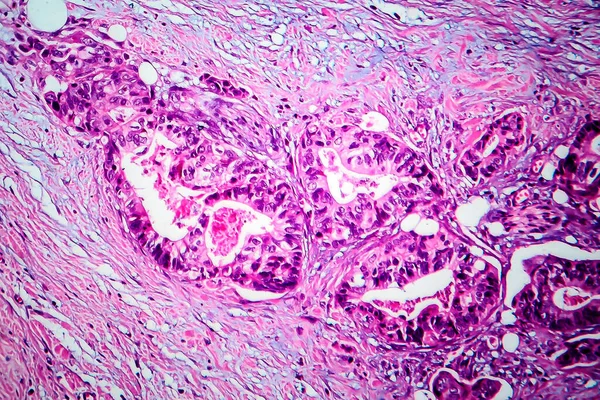
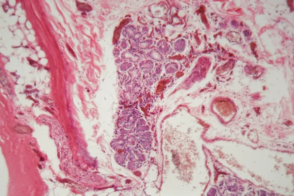
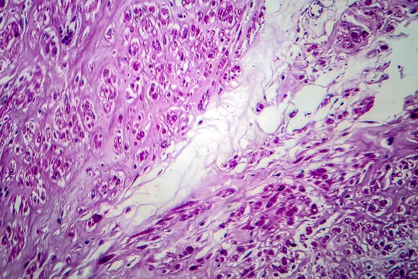
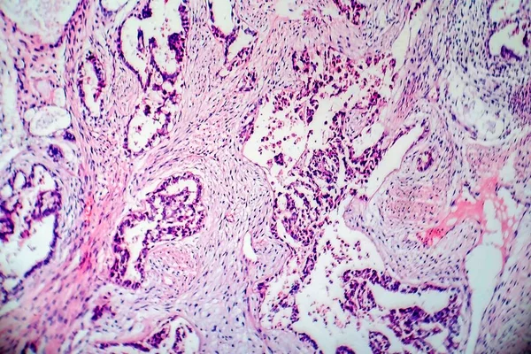
Stock image Light micrograph of teratoma, a tumor made up of several different types of tissue, such as hair, teeth, muscle, or bone. Teratoma is typically found in the ovary, testicle, or coccyx
Published:
Aug.27, 2021 14:22:52
Author:
katerynakon
Views:
0
Downloads:
0
File type:
image
/
jpg
File size:
8.87 MB
Orginal size:
4182
x
2788
px
Available sizes:
S
(500 x 333)
M
(1000 x 667)
L
(2000 x 1333)
XL
(4182 x 2788)
EL0
(4182 x 2788)
Level:
silver
Download Image
Image topics:
White
Background
Circle
Human
Beauty
Detail
Micro
Health
Light
Blood
Medicine
Medical
Pink
Cell
Education
Sample
Biology
Lab
Laboratory
Science
Cancer
Research
Slide
Diagnosis
Germ
Microscope
Microbiology
Immature
Smear
Tumor
Testicle
Gynecology
Staining
Ovary
Cyst
Oncology
Hematology
Coccyx
Histology
Selective Focus
Close Up
Dermoid
Eosin
Hematoxylin
Histopathology
Teratoma
Similar stock images
Breast Ductal Carcinoma, Light Micrograph, Photo Under Microscope
4103 × 2735
Meningioma, A Slow-growing Tumor That Forms From The Meninges, Light Micrograph, Photo Under Microscope
4364 × 2909
Endometrial Adenocarcinoma, Light Micrograph, Photo Under Microscope
4428 × 2952
Papillary Thyroid Carcinoma, Light Micrograph, Photo Under Microscope. The Most Common Type Of Thyroid Cancer
4126 × 2751
Breast Cancer, Light Micrograph, Photo Under Microscope
4438 × 2959
Human Lung Tissue With Pulmonary Embolism Under A Microscope.
5616 × 3744
Breast Ductal Carcinoma, Light Micrograph, Photo Under Microscope
4103 × 2735
Uterine Leiomyoma, Also Known As Fibroids,a Benign Smooth Muscle Tumor Of The Uterus, Light Micrograph, Photo Under Microscope
4136 × 2757