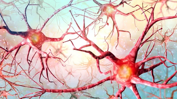
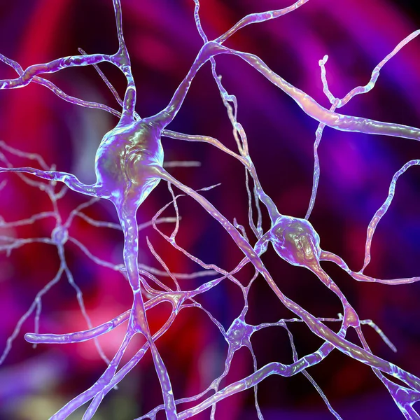
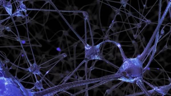
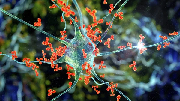
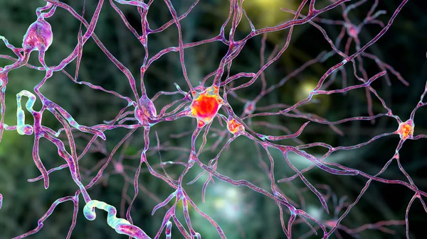
Stock image Intranuclear neuronal inclusions, 3D illustration. Intranuclear inclusions in neurons are found in different neurodegenerative diseases, including Huntingon's disease, spinocerebellar ataxia and other

Published: May.13, 2021 13:17:15
Author: katerynakon
Views: 45
Downloads: 12
File type: image / jpg
File size: 9.43 MB
Orginal size: 7200 x 4050 px
Available sizes:
Level: silver