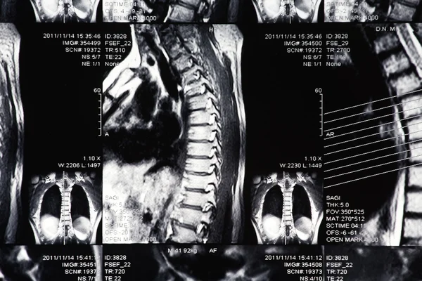
Stock image MRI C-Spine,neck, image 6view show Cervical neck dislocation.

Published: Apr.02, 2018 07:40:15
Author: Richmanphoto
Views: 74
Downloads: 1
File type: image / jpg
File size: 4.47 MB
Orginal size: 6000 x 4000 px
Available sizes:
Level: bronze