Home
Images
Vectors
Videos
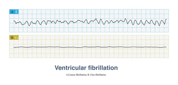
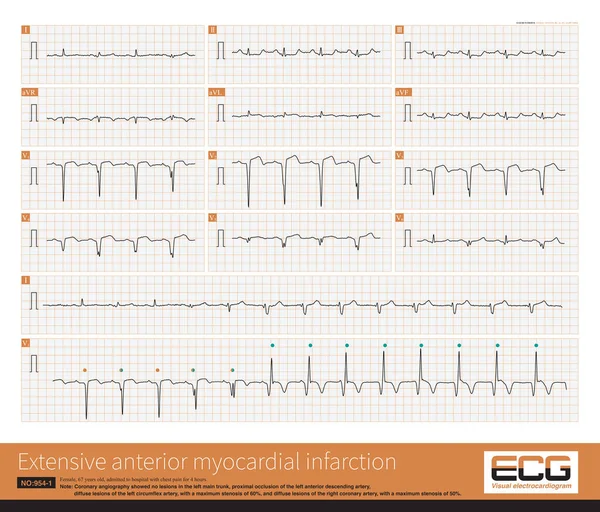
Stock image Male, 60 years old, clinically diagnosed as acute extensive anterior wall myocardial infarction. The patient died of ventricular fibrillation after admission.
Published:
Jul.04, 2022 07:53:07
Author:
asia11m
Views:
15
Downloads:
1
File type:
image
/
jpg
File size:
10.58 MB
Orginal size:
10000
x
6427
px
Available sizes:
S
(500 x 321)
M
(1000 x 643)
L
(2000 x 1285)
XL
(10000 x 6427)
EL0
(10000 x 6427)
Level:
beginner
Download Image
Image topics:
White
Blue
Background
Graphic
Illustration
Isolated
Human
Health
Medicine
Medical
Care
Technology
Line
Square
Wave
Cardiology
Heart
Monitor
Pulse
Letter
Treatment
Science
Death
Graph
Beat
Rhythm
Diagnosis
Heartbeat
Cardiogram
Cardiac
Ekg
Electrocardiogram
Ecg
Arrhythmia
Chest Pain
Cardiopulmonary Resuscitation
Cardiac Arrest
Myocardial Infarction
Coronary Heart Disease
Sudden Death
Myocardial Ischemia
Acute Coronary Syndrome
Stable Angina
Ecg Education
Qrs Wave
T Wave Inversion
Death Ecg
Similar stock images
Male, 60 Years Old, Clinically Diagnosed As Acute Extensive Anterior Wall Myocardial Infarction. The Patient Died Of Ventricular Fibrillation After Admission.
10000 × 4656
Tachy-brady Syndrome Is A Special Type Of In Which Bradycardia And Tachycardia Are Repeated Alternating Episodes.This ECG Recorded Episodes Of Paroxysmal Atrial Fibrillation.
10000 × 7447
Ventricular Fibrillation Is A Fatal Arrhythmia And Also A Cardiac Arrest Rhythm. It Can Be Divided Into Coarse Fibrillation And Fine Fibrillation According To The Amplitude Of The Fibrillation Wave.
10000 × 4784
A Patient With Extensive Anterior Myocardial Infarction Had Wide QRS Wave Tachycardia, That Is, Persistent Ventricular Tachycardia, With Atrioventricular Separation.
10000 × 8171
Ventricular Premature Contractions In Late Diastole Can Be Combined With Sinus Impulses To Excite The Ventricles And Produce Ventricular Fusion Waves.
10000 × 10641
A Fascicular Ventricular Tachycardia Is An Idiopathic Ventricular Tachycardia That Originates In The Left Fascicular Region, In This Case The Left Posterior Fascicular Region.
10000 × 9424
The Culprit Vessel Of Extensive Anterior Wall Ventricular Infarction Is The Most Common Proximal Segment Of The Left Anterior Descending Artery, Followed By The Middle-distal Segment.
10000 × 8549
The Depolarization Time Of The Right Atrium Is Prolonged, Causing The Co-depolarization Time Of The Left And Right Atria To Be Prolonged, So The P Wave Amplitude Increases.
10000 × 6929