
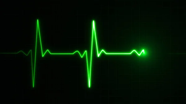
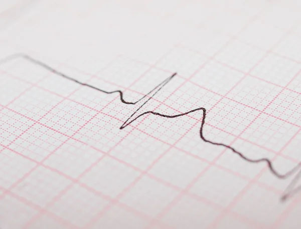
Stock image Death Ecg
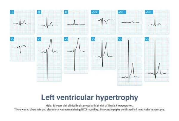
Sometimes, Left Ventricular Hypertrophy With Tall T Waves Is Easily Misdiagnosed As Hyperkalemia And Hyperacute T Waves, And ECG Needs To Be Carefully Identified In Combination With Clinic.
Image, 13.77MB, 10000 × 6782 jpg

Torsade De Pointes Refers To The Pleomorphic Ventricular Tachycardia That Occurs In The Background Of Long QT Interval, And The Polarity Of QRS Wave Twists Around The Equipotential Line.
Image, 18.28MB, 10000 × 5808 jpg


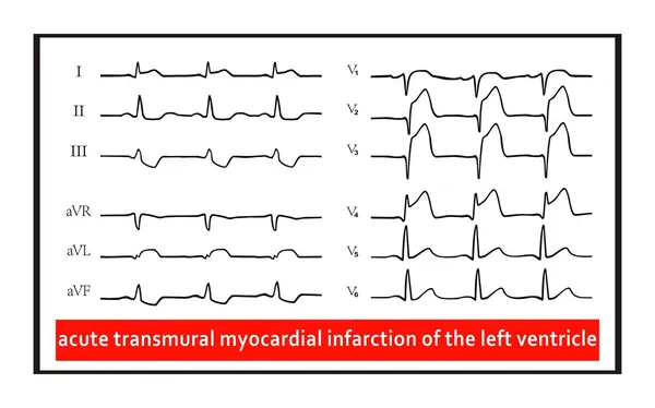
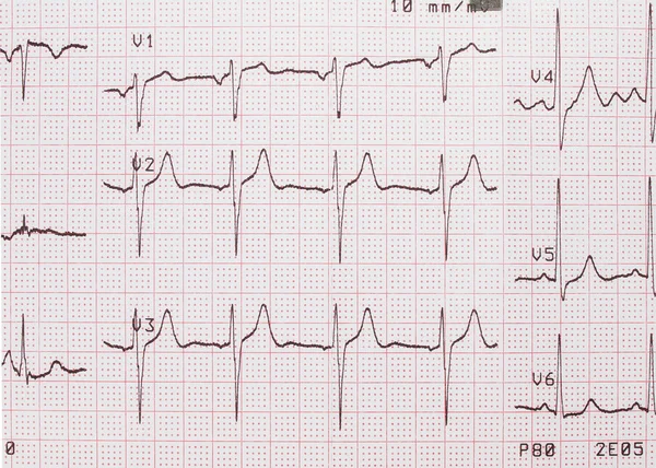
Male, 60 Years Old, Clinically Diagnosed As Acute Extensive Anterior Wall Myocardial Infarction. The Patient Died Of Ventricular Fibrillation After Admission.
Image, 10.58MB, 10000 × 6427 jpg

Male, 60 Years Old, Clinically Diagnosed As Acute Extensive Anterior Wall Myocardial Infarction. The Patient Died Of Ventricular Fibrillation After Admission.
Image, 6.05MB, 10000 × 4656 jpg
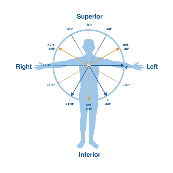
In The Frontal Lead System, The Lead Axes Of The 6 Limb Leads Form A Hexaxial Reference System, Which Is One Of The Important Theories Of Electrocardiography.
Image, 8.21MB, 10000 × 10000 jpg

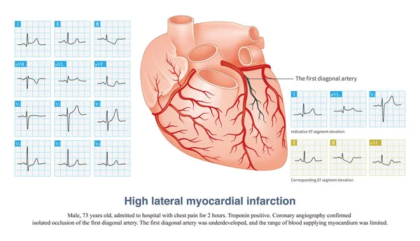
In Acute High Lateral Myocardial Infarction, There Is Indicative ST Segment Elevation In Leads I And AVL, And Corresponding ST Segment Depression In Leads II, III And AVF.
Image, 12.63MB, 10000 × 5739 jpg

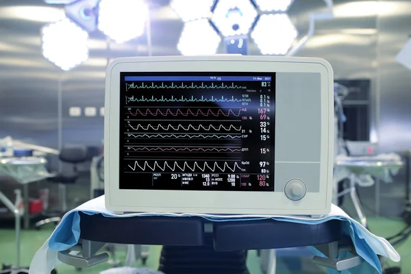


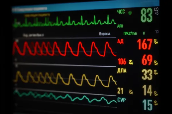
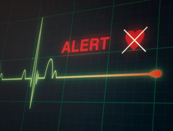


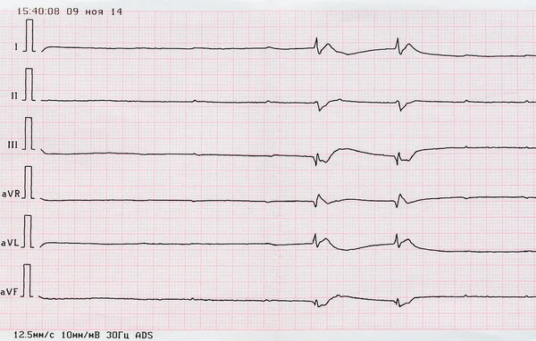
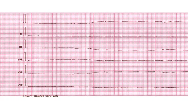
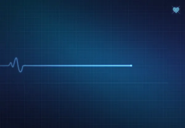
Emergency Cardiology And Resuscitation. ECG With Ventricular Asystole
Image, 4.44MB, 2678 × 1550 jpg


Electromechanical Separation Is A Kind Of Terminal ECG. The Patient's ECG Has Electrical Signals, The ECG Wave Is Widened With Morphological Abnormalities, And The Ventricle Has No Contraction.
Image, 21.68MB, 10000 × 10515 jpg
Page 1 >> Next