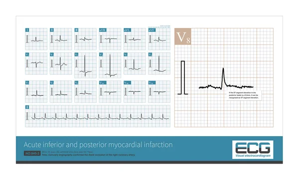
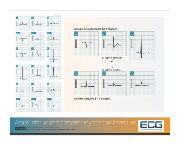
Stock image Male, 68 years old, chest pain for 7 hours. Coronary angiography suggests occlusion of the distal right coronary artery. The patient was successfully placed with a coronary stent.
Published: Jun.13, 2023 11:28:38
Author: asia11m
Views: 3
Downloads: 0
File type: image / jpg
File size: 15.13 MB
Orginal size: 10000 x 8095 px
Available sizes:
Level: beginner