photo free
Home Images Vectors Videos

Stock image Left Ventricle

stock vector Ventricular septal defect (VSD).Congenital heart defect

Ventricular Septal Defect (VSD).Congenital Heart Defect

Vector, 10.77MB, 5001 × 3334 eps
stock image Human Heart Anatomy

Human Heart Anatomy

Image, 3.15MB, 3840 × 2160 jpg
stock image Human Heart Anatomy

Human Heart Anatomy

Image, 3.71MB, 3840 × 2160 jpg
stock image Human Heart

Human Heart

Image, 2.71MB, 4096 × 4096 jpg
stock image Catheterization and small red heart. Cardiac ventriculography is a medical imaging test used to determine a patient cardiac function in the right or left ventricle

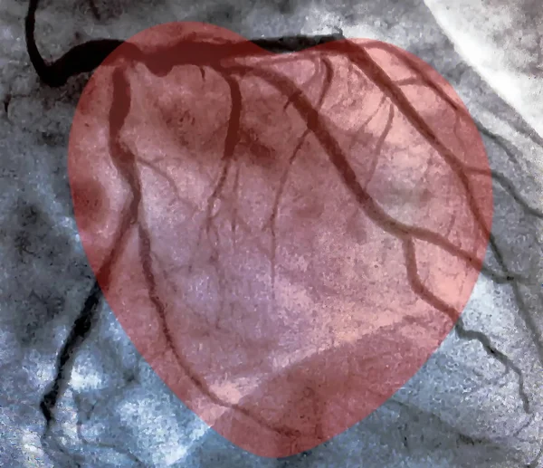
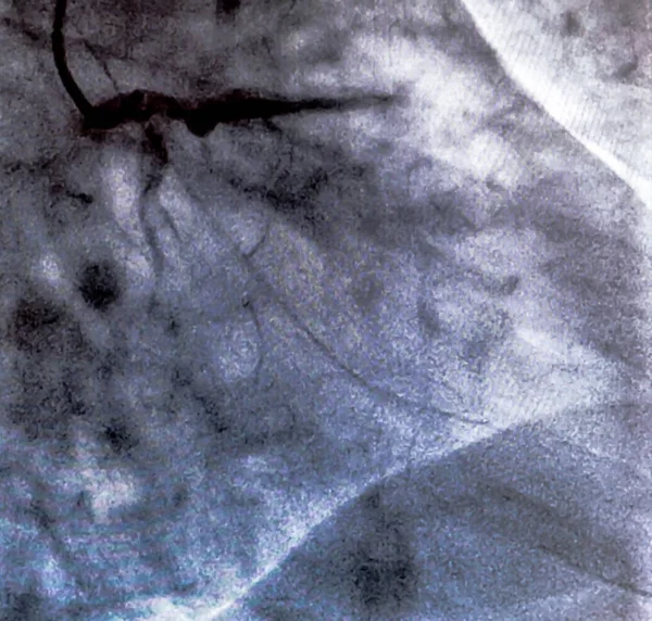
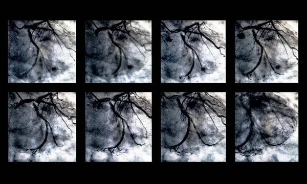
Catheterization And Small Red Heart. Cardiac Ventriculography Is A Medical Imaging Test Used To Determine A Patient Cardiac Function In The Right Or Left Ventricle

Image, 1.12MB, 3013 × 2600 jpg
stock image Catheterization. Cardiac ventriculography is a medical imaging test used to determine a patient cardiac function in the right or left ventricle

Catheterization. Cardiac Ventriculography Is A Medical Imaging Test Used To Determine A Patient Cardiac Function In The Right Or Left Ventricle

Image, 1.4MB, 3028 × 2890 jpg
stock image Ventricular tachyarrhythmia includes many clinical types, some benign and some malignant. For malignant ventricular arrhythmias, patients are at risk of death.

Ventricular Tachyarrhythmia Includes Many Clinical Types, Some Benign And Some Malignant. For Malignant Ventricular Arrhythmias, Patients Are At Risk Of Death.

Image, 27.66MB, 8000 × 10973 jpg
stock image Human Heart Disease Therapy

Human Heart Disease Therapy

Image, 5.94MB, 4150 × 4144 jpg
stock image Catheterization. Cardiac ventriculography is a medical imaging test used to determine a patient cardiac function in the right or left ventricle

Catheterization. Cardiac Ventriculography Is A Medical Imaging Test Used To Determine A Patient Cardiac Function In The Right Or Left Ventricle

Image, 2.42MB, 3007 × 2930 jpg
stock vector simple vector illustration of the circulatory system focused on the heart and lungs with the names of each part written in English on a black background.

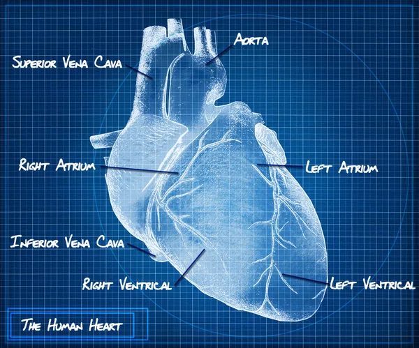
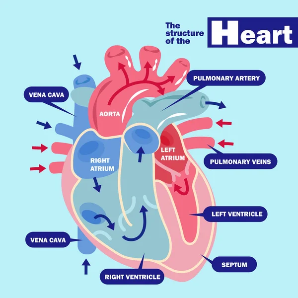
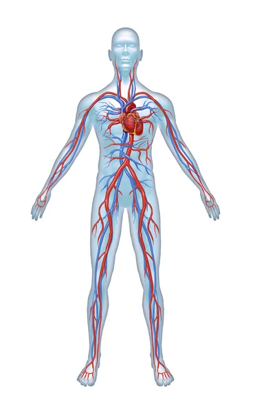
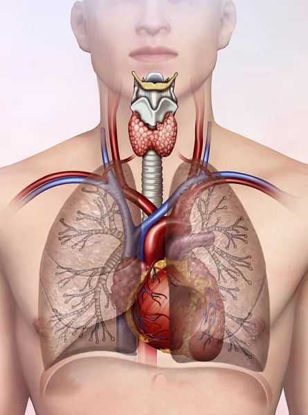
Simple Vector Illustration Of The Circulatory System Focused On The Heart And Lungs With The Names Of Each Part Written In English On A Black Background.

Vector, 0.98MB, 4800 × 3600 eps
stock image Human heart concept anatomy on a grunge background as a medical health care symbol  or cardiology icon of an inner cardiovascular organ in a 3D illustration style.

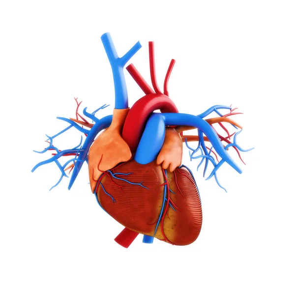
Human Heart Concept Anatomy On A Grunge Background As A Medical Health Care Symbol Or Cardiology Icon Of An Inner Cardiovascular Organ In A 3D Illustration Style.

Image, 13.86MB, 5305 × 3393 jpg
stock vector Anti Valentines day banner,  My heart beats for you. vector illustration

Anti Valentines Day Banner, My Heart Beats For You. Vector Illustration

Vector, 15.79MB, 3751 × 6542 eps
stock image The Human heart blueprint concept.

The Human Heart Blueprint Concept.

Image, 24.56MB, 5000 × 4167 jpg
stock vector Anti valentine's vintage card, vector illustration

Anti Valentine's Vintage Card, Vector Illustration

Vector, 7.48MB, 4168 × 6084 eps
stock vector Human Heart Cross Section

Human Heart Cross Section

Vector, 0.31MB, 4657 × 4657 eps
stock vector Anti Valentines day card,  Skeleton holding a flower rose, vector illustration

Anti Valentines Day Card, Skeleton Holding A Flower Rose, Vector Illustration

Vector, 4.39MB, 4167 × 4707 eps
stock image Human Heart Puzzle

Human Heart Puzzle

Image, 2.89MB, 2590 × 2685 jpg
stock image Female, 51 years old, diagnosed with mitral stenosis. When this ECG was taken, the patient still maintained sinus rhythm.Note that the P wave duration was widened.

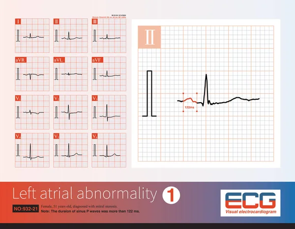
Female, 51 Years Old, Diagnosed With Mitral Stenosis. When This ECG Was Taken, The Patient Still Maintained Sinus Rhythm.Note That The P Wave Duration Was Widened.

Image, 14.21MB, 10000 × 7772 jpg
stock vector Anti Valentine's day,  hands hold a heart. vector illustration

Anti Valentine's Day, Hands Hold A Heart. Vector Illustration

Vector, 14.86MB, 2362 × 3829 eps
stock image Heart disease awareness and cardiovascular illness research and stroke prevention as a medical health symbol for high blood pressure hypertension and Arrhythmia with 3D illustration elements

Heart Disease Awareness And Cardiovascular Illness Research And Stroke Prevention As A Medical Health Symbol For High Blood Pressure Hypertension And Arrhythmia With 3D Illustration Elements

Image, 7.56MB, 7655 × 3500 jpg
stock image Heart Pain symbol

Heart Pain Symbol

Image, 4.07MB, 3342 × 3342 jpg
stock vector Human heart

Human Heart

Vector, 19.03MB, 5417 × 3325 eps
stock image Catheterization. Cardiac ventriculography is a medical imaging test used to determine a patient cardiac function in the right or left ventricle

Catheterization. Cardiac Ventriculography Is A Medical Imaging Test Used To Determine A Patient Cardiac Function In The Right Or Left Ventricle

Image, 2.14MB, 2620 × 2500 jpg
stock vector Watercolor seamless pattern with realistic human heart on the white background, aquarelle.  Vector illustration.

Watercolor Seamless Pattern With Realistic Human Heart On The White Background, Aquarelle. Vector Illustration.

Vector, 12.35MB, 5002 × 5003 eps
stock vector heart with health concept

Heart With Health Concept

Vector, 2.52MB, 6000 × 6000 eps
stock vector Watercolor realistic human heart on the white background, aquarelle.  Vector illustration. Hand-drawn decorative element useful for invitations, scrapbooking, design.

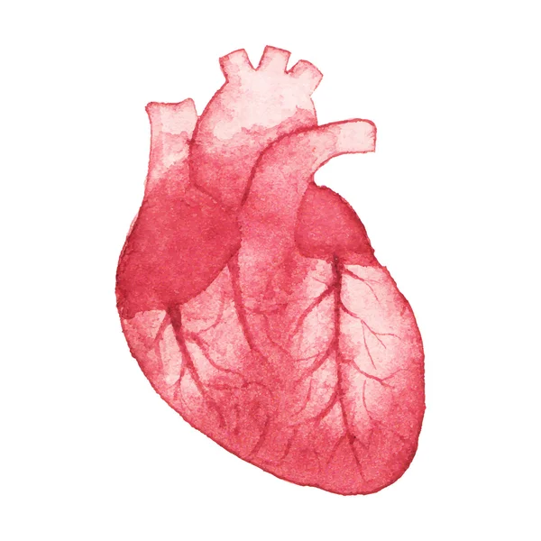
Watercolor Realistic Human Heart On The White Background, Aquarelle. Vector Illustration. Hand-drawn Decorative Element Useful For Invitations, Scrapbooking, Design.

Vector, 10.31MB, 5000 × 5000 eps
stock image Structure of Human Heart. Black and white illustration (outline).

Structure Of Human Heart. Black And White Illustration (outline).

Image, 3.16MB, 9375 × 11458 jpg
stock image Catheterization. Cardiac ventriculography is a medical imaging test used to determine a patient cardiac function in the right or left ventricle

Catheterization. Cardiac Ventriculography Is A Medical Imaging Test Used To Determine A Patient Cardiac Function In The Right Or Left Ventricle

Image, 3.08MB, 3333 × 2000 jpg
stock image In acute myocardial ischemia, the amplitude of T wave is increased first, and then the ST segment is elevated. When the end of QRS wave is deformed,  there is a lack of collateral circulation.

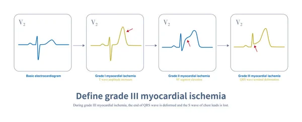
In Acute Myocardial Ischemia, The Amplitude Of T Wave Is Increased First, And Then The ST Segment Is Elevated. When The End Of QRS Wave Is Deformed, There Is A Lack Of Collateral Circulation.

Image, 1.32MB, 10108 × 4093 jpg
stock image Healthy Heart Food

Healthy Heart Food

Image, 10.03MB, 6371 × 4500 jpg
stock vector Aorta largest human artery structure vertebral column diagram hand drawn schematic vector illustration. Medical science educational illustration

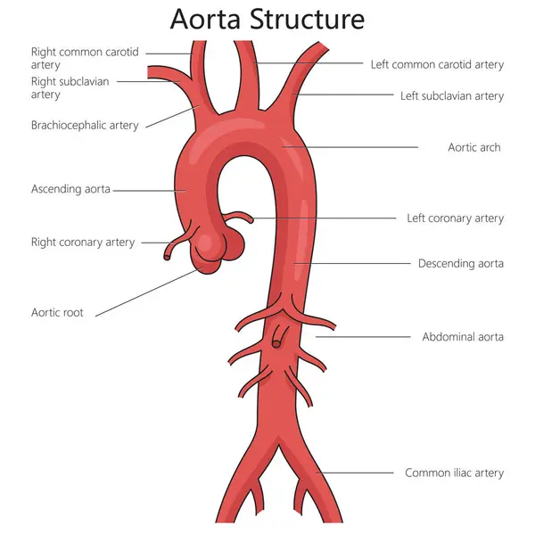
Aorta Largest Human Artery Structure Vertebral Column Diagram Hand Drawn Schematic Vector Illustration. Medical Science Educational Illustration

Vector, 0.61MB, 4000 × 4000 eps
stock image Viral myocarditis or virus infection of the human heart resulting in inflammation  of the cardiac circulatory organ with 3D illustration elements.

Viral Myocarditis Or Virus Infection Of The Human Heart Resulting In Inflammation Of The Cardiac Circulatory Organ With 3D Illustration Elements.

Image, 20.08MB, 7085 × 3506 jpg
stock image Generally, when the sinus heart rate is below 60 beats per minute, it is called sinus bradycardia. This arrhythmia can be both physiological and often pathological.

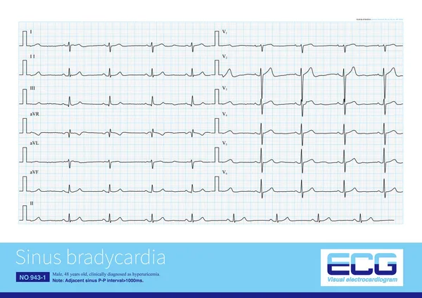
Generally, When The Sinus Heart Rate Is Below 60 Beats Per Minute, It Is Called Sinus Bradycardia. This Arrhythmia Can Be Both Physiological And Often Pathological.

Image, 20.38MB, 10000 × 7069 jpg
stock vector Abstrack Human heart anatomy

Abstrack Human Heart Anatomy

Vector, 2.17MB, 5586 × 7930 eps
stock image Atrial focal originating in the left upper pulmonary vein, with an upright P wave in V1 and wide duration,  inverted P wave in lead aVL and an upright P wave with notch in inferior leads.

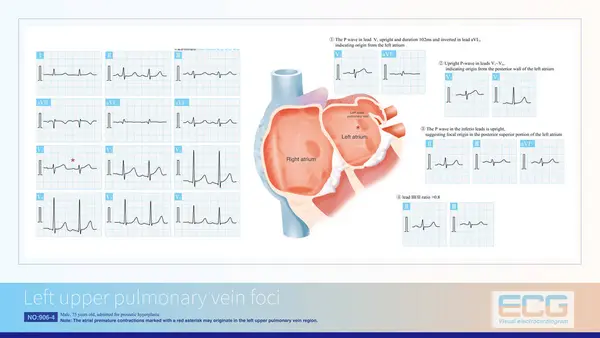
Atrial Focal Originating In The Left Upper Pulmonary Vein, With An Upright P Wave In V1 And Wide Duration, Inverted P Wave In Lead AVL And An Upright P Wave With Notch In Inferior Leads.

Image, 12.65MB, 10000 × 5632 jpg
stock image This is a pathological photo of human left ventricular hypertrophy, showing an increase in myocardial diameter and interstitial distance.Magnify 40x.

This Is A Pathological Photo Of Human Left Ventricular Hypertrophy, Showing An Increase In Myocardial Diameter And Interstitial Distance.Magnify 40x.

Image, 42.86MB, 8500 × 8500 jpg
stock image Obstructed vein due to thrombosis and normal vein with blood cells. 3D rendering

Obstructed Vein Due To Thrombosis And Normal Vein With Blood Cells. 3D Rendering

Image, 6.72MB, 5000 × 4552 jpg
stock vector Medical illustration that compares a normal heart with hearts afflicted by ventricular septal defects, an abnormal opening (hole) in the heart, with annotations.

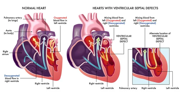
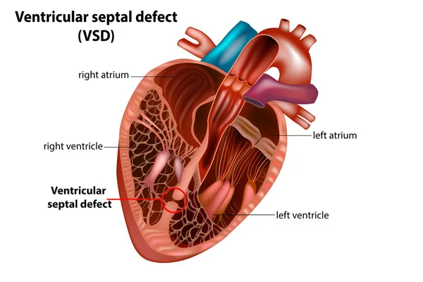
Medical Illustration That Compares A Normal Heart With Hearts Afflicted By Ventricular Septal Defects, An Abnormal Opening (hole) In The Heart, With Annotations.

Vector, 9.41MB, 7000 × 3686 eps
stock image Human Heart

Human Heart

Image, 1.75MB, 2890 × 3006 jpg
stock image The typical ST-T changing of left ventricular hypertrophy are:  ST segment slightly convex with downward sloping depression; fusion of ST segment and inverted T wave;  asymmetry of inverted T wave.

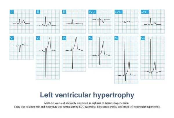
The Typical ST-T Changing Of Left Ventricular Hypertrophy Are: ST Segment Slightly Convex With Downward Sloping Depression; Fusion Of ST Segment And Inverted T Wave; Asymmetry Of Inverted T Wave.

Image, 11.52MB, 10000 × 8453 jpg
stock image Male, 65 years old, was clinically diagnosed with acute anterior myocardial infarction. The patient was treated with a coronary stent, but no reperfusion T wave occurred on day 2.

Male, 65 Years Old, Was Clinically Diagnosed With Acute Anterior Myocardial Infarction. The Patient Was Treated With A Coronary Stent, But No Reperfusion T Wave Occurred On Day 2.

Image, 19.72MB, 10000 × 8695 jpg
stock vector A typical representation of the section of the stomach, with the parts, 1: Right Auricle; 2: Left Auricle; 3: Right Ventricle; 4: Left Ventricle; 5: Systemic Veins; 6: Pulmonary Veins; 7: Aorta; and other, vintage line drawing or engraving illustrati

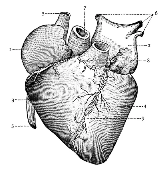
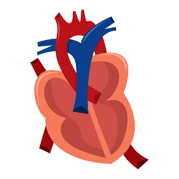
A Typical Representation Of The Section Of The Stomach, With The Parts, 1: Right Auricle; 2: Left Auricle; 3: Right Ventricle; 4: Left Ventricle; 5: Systemic Veins; 6: Pulmonary Veins; 7: Aorta; And Other, Vintage Line Drawing Or Engraving Illustrati

Vector, 5.42MB, 9491 × 9718 eps
stock vector Heart Anatomy diagram. Human Heart Structure. Labelled heart half in doodle style. Part of heart foe education. Hand drew vector illustration.

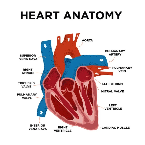
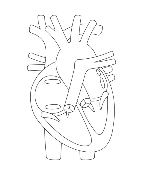
Heart Anatomy Diagram. Human Heart Structure. Labelled Heart Half In Doodle Style. Part Of Heart Foe Education. Hand Drew Vector Illustration.

Vector, 9.06MB, 5000 × 5000 eps
stock image Sometimes, left ventricular hypertrophy with tall T waves is easily misdiagnosed as hyperkalemia and hyperacute T waves, and ECG needs to be carefully identified in combination with clinic.

Sometimes, Left Ventricular Hypertrophy With Tall T Waves Is Easily Misdiagnosed As Hyperkalemia And Hyperacute T Waves, And ECG Needs To Be Carefully Identified In Combination With Clinic.

Image, 13.77MB, 10000 × 6782 jpg
stock image A synthetic graft is an artificial tube that allows the oxygenated blood to flowfrom the heart to the rest of the body. 3D rendering

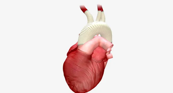
A Synthetic Graft Is An Artificial Tube That Allows The Oxygenated Blood To Flowfrom The Heart To The Rest Of The Body. 3D Rendering

Image, 3.17MB, 7258 × 3920 jpg
stock image Human Cardiovascular System

Human Cardiovascular System

Image, 1.78MB, 2310 × 3761 jpg
stock image Human heart

Human Heart

Image, 6.02MB, 6200 × 6200 jpg
stock image In acute left main occlusion, the left ventricular myocardium is massively ischemic and necrotic, the excitatory potential of the left ventricle is weakened, and the axis may deviate to the right .

In Acute Left Main Occlusion, The Left Ventricular Myocardium Is Massively Ischemic And Necrotic, The Excitatory Potential Of The Left Ventricle Is Weakened, And The Axis May Deviate To The Right .

Image, 12.47MB, 10000 × 6364 jpg
stock image Male, 65 years old, was clinically diagnosed with acute anterior myocardial infarction. The patient was treated with a coronary stent, but no reperfusion T wave occurred on day 2.

Male, 65 Years Old, Was Clinically Diagnosed With Acute Anterior Myocardial Infarction. The Patient Was Treated With A Coronary Stent, But No Reperfusion T Wave Occurred On Day 2.

Image, 25.24MB, 17956 × 5906 jpg
stock image Human respiratory cardio system

Human Respiratory Cardio System

Image, 5.06MB, 2741 × 3696 jpg
Page 1 >> Next