
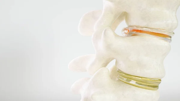
Stock image Nerve Root
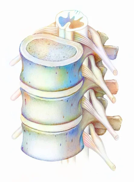
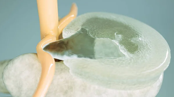


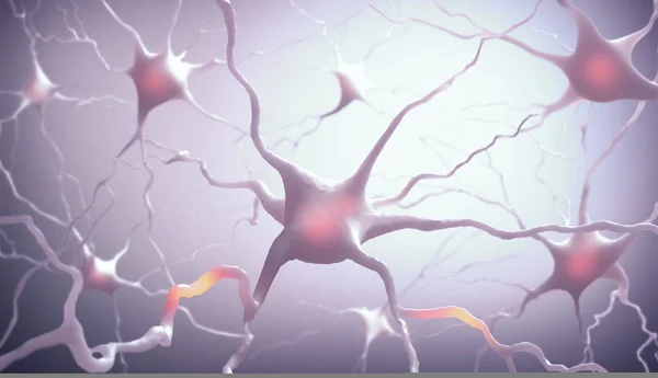


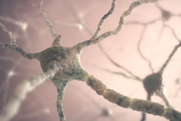
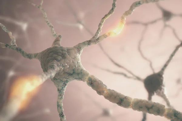


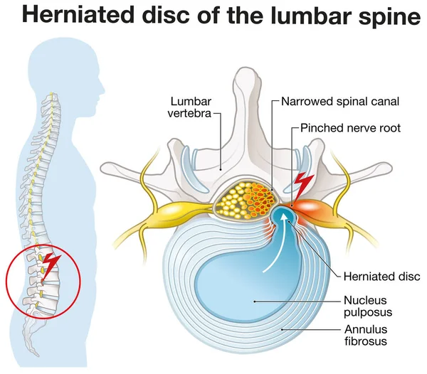
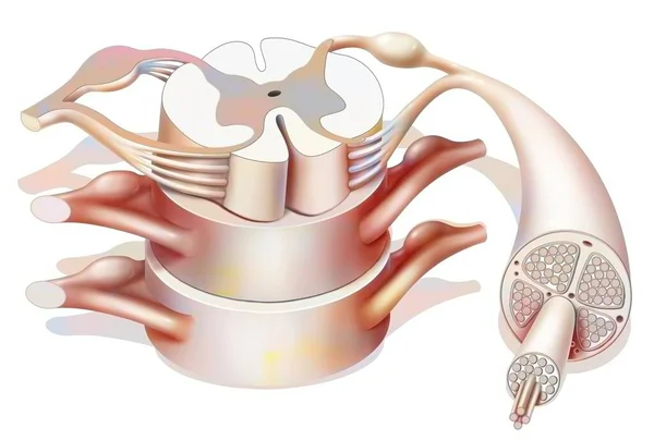
Illustration Showing Lumbar Vertebra With Intervertebral Disc And Herniated Nucleus Pulposus
Image, 3.81MB, 4870 × 4273 jpg

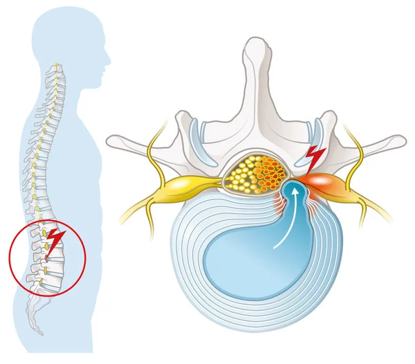
Illustration Showing Lumbar Vertebra With Intervertebral Disc And Herniated Nucleus Pulposus
Image, 3.56MB, 4870 × 4273 jpg

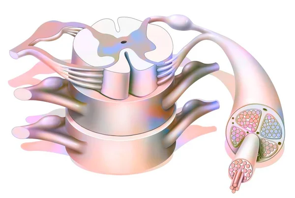
Illustration Showing Healthy Lumbar Vertebrae And Intervertebral Disc. Labeled Illustration
Image, 2.38MB, 5000 × 3208 jpg
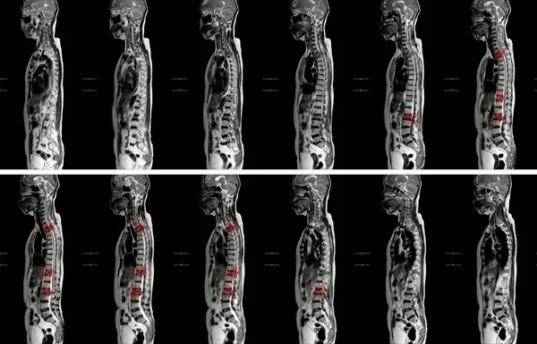
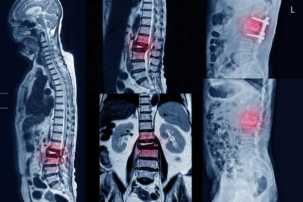
MRI OF THORACOLUMBAR SPINE IMPRESSION: Moderate Pathological Compression Of T11 And L2 Levels With Enhancing Multiple Marrow Lesions At T1, T10 ToT12, L2, L3 To L5 Levels.
Image, 4.96MB, 5688 × 3643 jpg
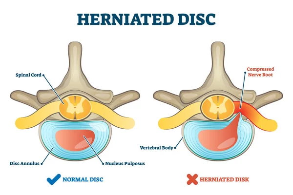
Herniated Disc Injury As Labeled Spinal Pain Explanation Vector Illustration
Vector, 7.09MB, 5000 × 3333 eps

Illustration Showing Healthy Lumbar Vertebrae. Different Views. Labeled Illustration
Image, 3.82MB, 6000 × 4806 jpg
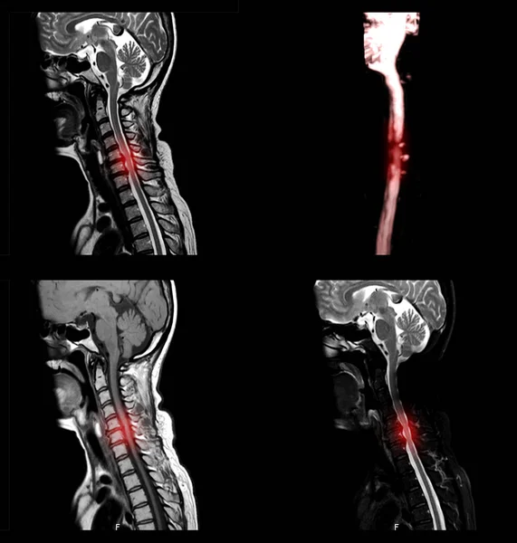
MRI C-spine A Female 67 Year Old.Impression Moderate Bening Compression Fracture At C4 And C5 Vertebrae.
Image, 3.99MB, 2860 × 2371 jpg
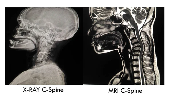
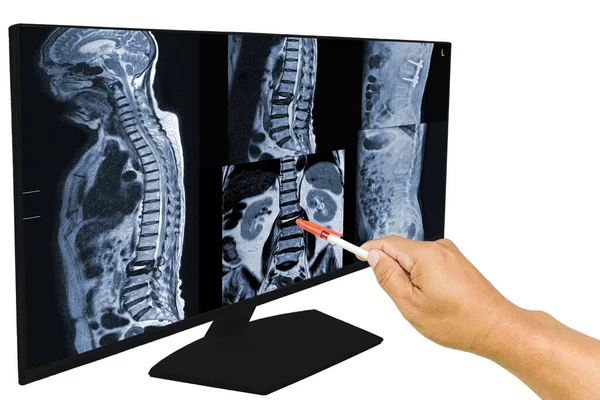
Cevical Spine Image Normal X-ray And MRI : Showing Severe Narrowing Disc Space C4-5 With Erosion And Sclerosis Of End Plates.
Image, 10.35MB, 6192 × 3561 jpg

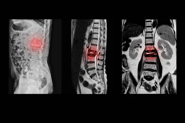
MRI Of Lumbar Spine The Study Reveals Burst Fracture Of L2 Vertebral Body, Appears As Severe Decreased Disc Height And Widening Of Interpedicular Distance.
Image, 2.77MB, 3096 × 4128 jpg
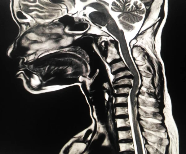
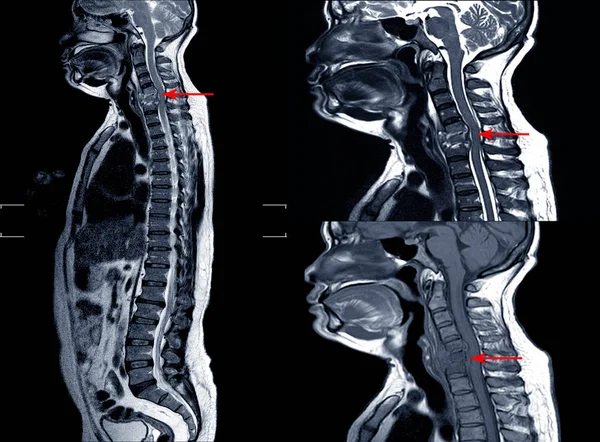
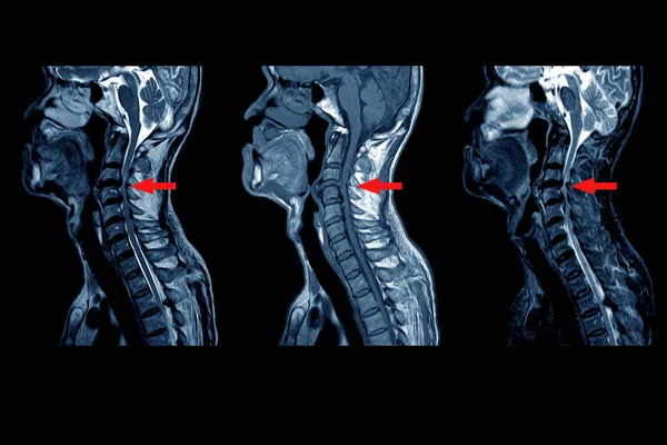
A Sagittal View Of MRI C-spine Or Magnetic Resonance Image Of Cervical Spine Showing Spondylosis Causing Cervical Spondylotic Myelopathy And Compression Fracture.
Image, 1.46MB, 2835 × 2976 jpg


Collection MRI Of Lumbar Spine History Of Fall With Back Pain, Radiate To Leg, Rule Out Spinal Stenosis .Impression:Burst Fracture Of L2 Vertebral Body With Severe Vertebral Collapse.Medical Concept.
Image, 8.31MB, 5820 × 3890 jpg
MRI OF CERVICAL SPINE : Moderate To Severe Posterior Central Disc Protrusion Of C3/4 To C5/6 Intervertebral Discs With A 2.0 Cm In Length Small Posterior Subligamentous Fluid Collection.on Red Point
Image, 4.36MB, 6000 × 4000 jpg

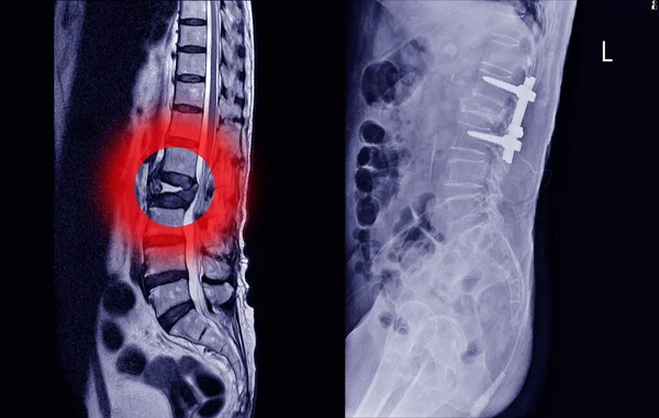
Medical X-ray And MRI Of Lumbar Spine Compression Fracture Bulging Of L1-2. On Arrow Point..Lumbar Spondylosis From L1-2 To L5-S1 Discs.Medical Healthcare Concept.
Image, 6.06MB, 5500 × 3500 jpg
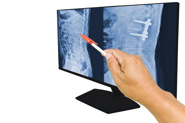
The Doctor Reported The MRI Scans Of The Lumbar Spine Compression Fracture Bulging Of L1-L2. And Post Operation Fixed By Iron Rod And Screws. Medical Education Concept.
Image, 6.16MB, 6000 × 4000 jpg

MRI Of Cervical Spine,Impression:Severe Compression Fracture Of C6 Vertebral Body Causing Kyphosis Of Cervical Spine, Too Soft And Blurry Image.
Image, 6.88MB, 6000 × 4000 jpg

Cardiologist Doctor Holding Stethoscopet, Blurred Electrocardiogram Result On Paper With AICD Pacemaker In Chest X-ray Background.medical Concept
Image, 5.46MB, 5500 × 3500 jpg

The Doctor Reported The MRI Scans Of The Lumbar Spine Compression Fracture Bulging Of L1-L2. And Post Operation Fixed By Iron Rod And Screws. Medical Education Concept.
Image, 6.18MB, 6000 × 3792 jpg

Adult Woman Treats The Cervical Spine With The Help Of Magnetic Field Physiotherapy, Magnetism, White Background, Osteochondrosis
Image, 8.48MB, 5184 × 3456 jpg
MRI Of Lumbar Spine History Of Fall With Back Pain, Radiate To Leg, Rule Out Spinal Stenosis .Burst Fracture Of L2 Vertebral Body With Severe Vertebral Collapse.
Image, 4.67MB, 6000 × 4000 jpg

The Doctor Reported The MRI Scans Of The Lumbar Spine Compression Fracture Bulging Of L1-L2. And Post Operation Fixed By Iron Rod And Screws. Medical Education Concept.
Image, 7.51MB, 6000 × 4000 jpg


Physiotherapy Of The Cervical Spine With The Help Of Physiotherapy, A Magnet, Reducing Inflammation And Improving Blood Circulation, Copy Space
Image, 7.84MB, 5184 × 3456 jpg
Page 1 >> Next