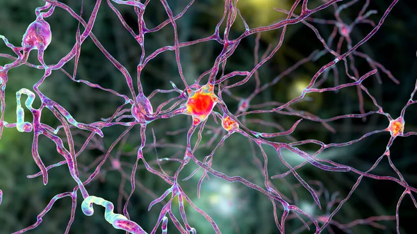
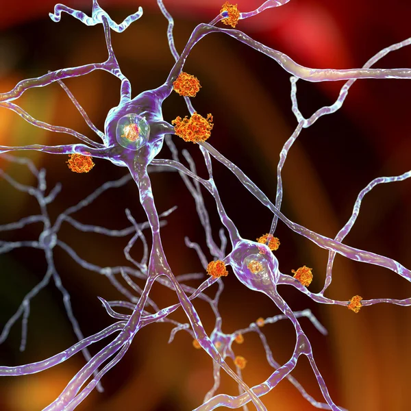
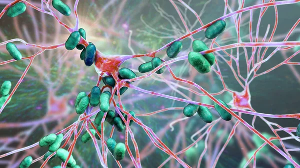
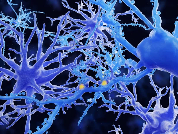
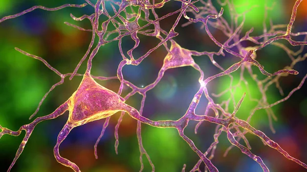
Stock image Neuronal inclusions in Huntington's disease, 3D illustration. Inclusions are composed of mutated huntingtin protein, they are initially formed at axons and dendrites, then migrate to nuclei of neurons

Published: May.13, 2021 13:17:15
Author: katerynakon
Views: 8
Downloads: 2
File type: image / jpg
File size: 9.3 MB
Orginal size: 7200 x 4050 px
Available sizes:
Level: silver