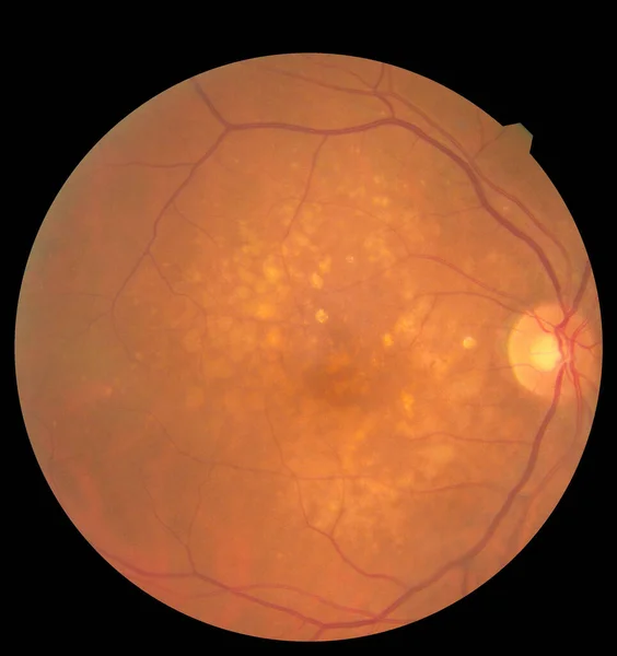
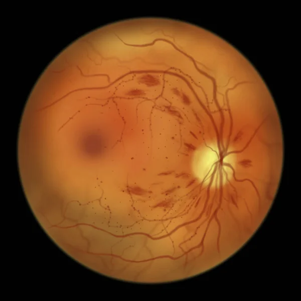
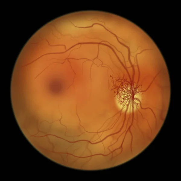
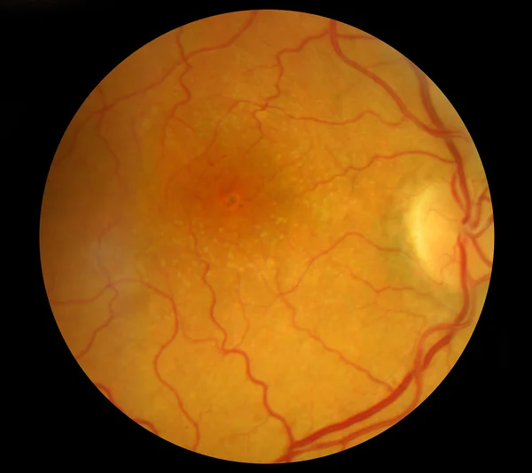
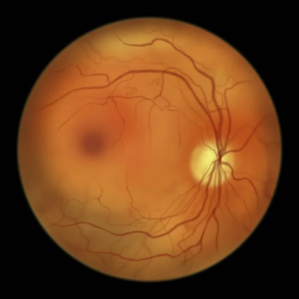
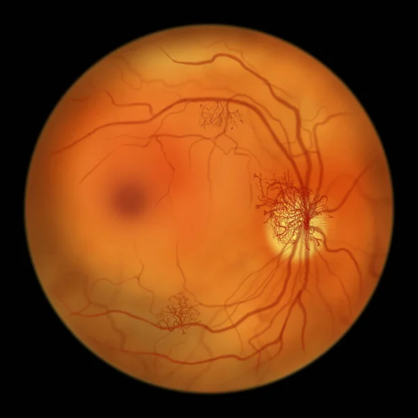
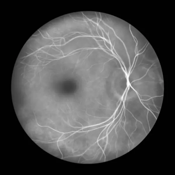
Stock image Normal retina, illustration of an ophthalmoscope image in fluorescein angiography.
Published: Sep.26, 2023 14:25:47
Author: katerynakon
Views: 2
Downloads: 0
File type: image / jpg
File size: 2.76 MB
Orginal size: 5000 x 5000 px
Available sizes:
Level: silver