Home
Images
Vectors
Videos
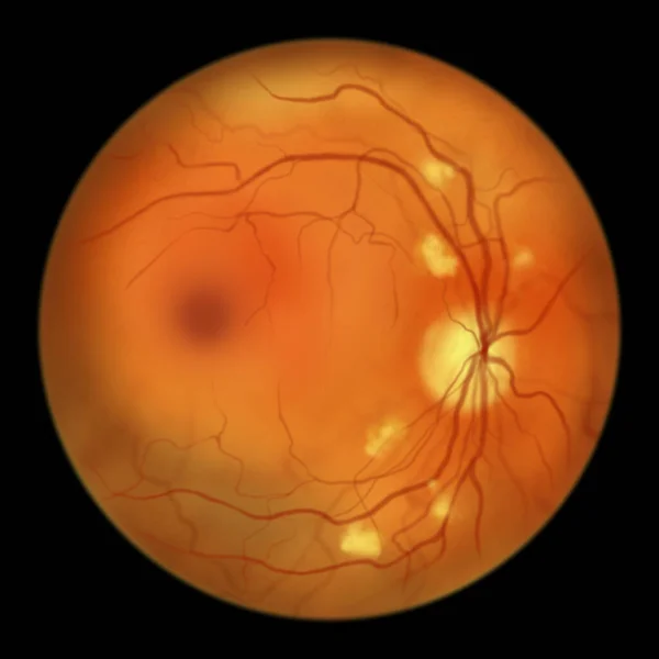
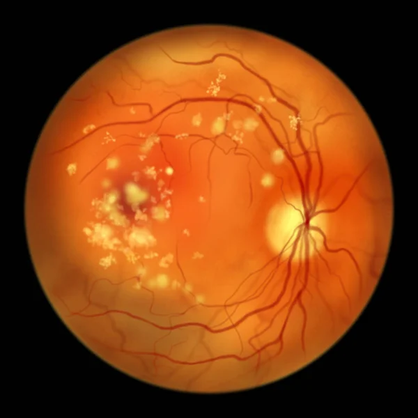
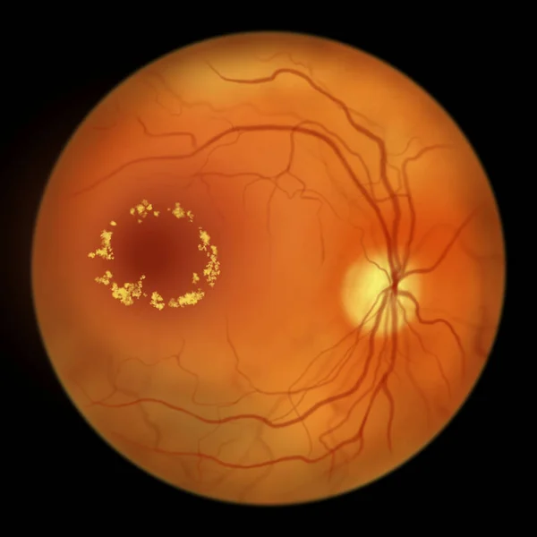
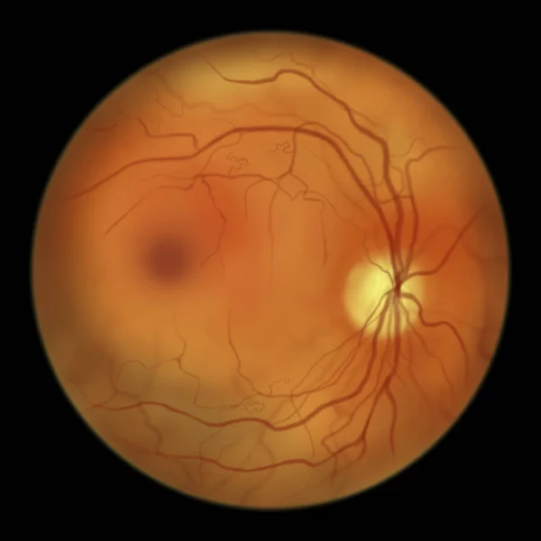
Stock image Retina in blastomycosis (infection caused by fungi Blastomyces dermatitidis) as seen during ophthalmoscopy. An illustration showing scattered yellow choroidal infiltrates and a choroidal mass lesion.
Published:
Nov.24, 2023 09:08:49
Author:
katerynakon
Views:
6
Downloads:
1
File type:
image
/
jpg
File size:
2.86 MB
Orginal size:
5000
x
5000
px
Available sizes:
S
(500 x 500)
M
(1000 x 1000)
L
(2000 x 2000)
XL
(5000 x 5000)
EL0
(5000 x 5000)
Level:
silver
Download Image
Image topics:
Illustration
Medical
Eye
Drawing
Disease
Optic
Disc
Fungi
Examination
Anatomy
Ophthalmic
Ocular
Ophthalmology
Chronic
Fungal
Microscopic
Vessels
Conditions
Abnormal
Diagnostics
Congenital
Vascular
Pathology
Retina
Nerve
Ophthalmoscopy
Retinal
Findings
Disorders
Infiltrate
Ophthalmoscope
Granuloma
Fundus
Black Background
Hand Drawn
Eye Examination
Medical Illustration
Optic Disc
Anomalies
Retinoscopy
Granulomatous
Abnormalities
Fundoscopy
Blastomycosis
Dermatitidis
Blastomyces
Retinal Anatomy
Tropical Infection
Similar stock images
Normal Retina, Illustration Of An Ophthalmoscope Image.
5000 × 5000
Non-proliferative Diabetic Retinopathy, Illustration Showing Cotton Wool Spots As Fluffy Yellow Patches, Abnormal Finding On Funduscopic Examination Of The Eye Retina In Diabetes Mellitus
5000 × 5000
Retinal Arteriovenous Malformation: Rare Congenital Retinal Vascular Anomalies With Tangled Blood Vessels In The Retina, Illustration Shows Artery-vein Communication Without Intervening Capillaries.
5000 × 5000
Roth Spots In The Retina As Observed During Ophthalmoscopy, An Illustration Showing White-centered Retinal Hemorrhages With Surrounding Hemorrhagic Rings.
5000 × 5000
Autosomal Recessive Bestrophinopathy, Ophthalmoscope View, Scientific Illustration Showing Accumulation Of Lipofuscin Deposits Around And Beyond The Macula Leading To Progressive Damage To The Retina
5000 × 5000
Diabetic Retinopathy, Illustration Showing Macula Edema And Hard Exudates, Abnormal Finding On Fundoscopic Examination Of The Eye Retina In Diabetes Mellitus
5000 × 5000
Non-proliferative Diabetic Retinopathy, Illustration Showing IRMAs (intraretinal Microvascular Abnormalities) As Small Vessels With Abnormal Branching Or Dilatation In Ischaemic Areas
5000 × 5000