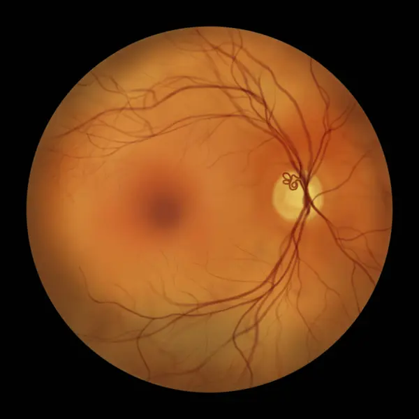
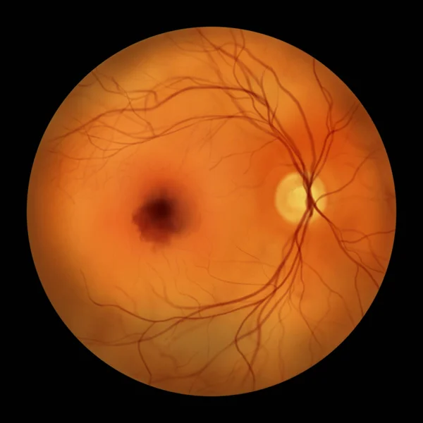
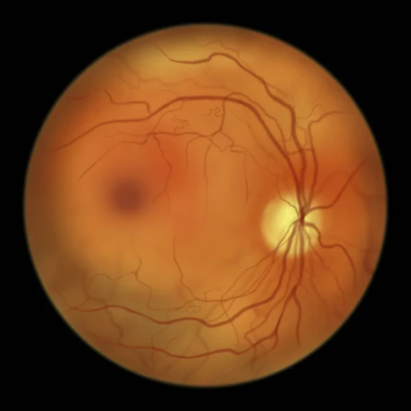
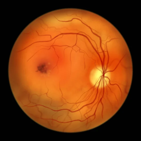
Stock image Retinal arteriovenous malformation: Rare congenital retinal vascular anomalies with tangled blood vessels in the retina, illustration shows artery-vein communication without intervening capillaries.

Published: Sep.26, 2023 14:24:42
Author: katerynakon
Views: 8
Downloads: 1
File type: image / jpg
File size: 2.64 MB
Orginal size: 5000 x 5000 px
Available sizes:
Level: silver