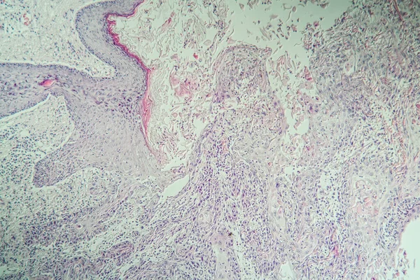
Stock image Spinal cord tissue section under the microscope 100x

Published: Apr.22, 2020 13:14:39
Author: dr.lange.unitybox.de
Views: 1
Downloads: 0
File type: image / jpg
File size: 18.98 MB
Orginal size: 5616 x 3744 px
Available sizes:
Level: bronze