Home
Images
Vectors
Videos
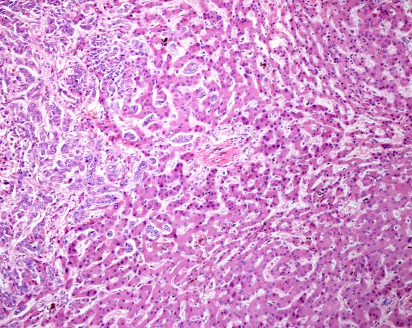
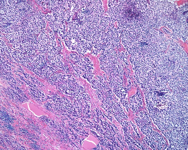
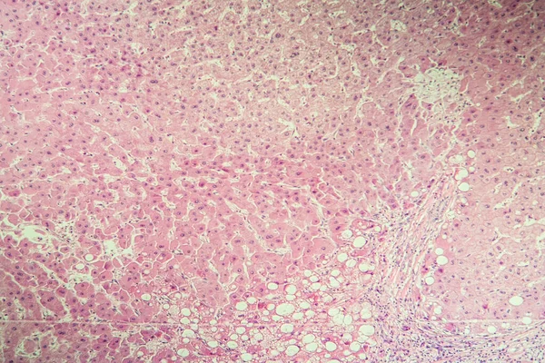
Stock image Human liver. Micronodular cirrhosis. Regenerating nodule of hepatocytes showing an extensive fatty change (steatosis), separated by fibrous septa with chronic inflammatory infiltrates.
Published:
Jul.04, 2022 16:30:27
Author:
jlcalvo@ucm.es
Views:
8
Downloads:
0
File type:
image
/
jpg
File size:
9.43 MB
Orginal size:
3840
x
3072
px
Available sizes:
S
(500 x 400)
M
(1000 x 800)
L
(2000 x 1600)
XL
(3840 x 3072)
EL0
(3840 x 3072)
Level:
beginner
Download Image
Image topics:
Healthy
Stain
Cellular
Fat
Biology
Alcohol
Cells
Tissue
Fatty
Anatomy
Obesity
Anatomical
Microscopy
Hepatic
Photomicrograph
Lipid
Histology
Histological
Hepatitis C
Hepatocyte
Fatty Liver
Histopathology
Liver Anatomy
Light Micrograph
Liver Cell
Light Microscope
Fat Deposit
Hepatic Lobule
Alcoholic Cirrhosis
Liver Lobule
Similar stock images
Papillary Thyroid Carcinoma, Light Micrograph, Photo Under Microscope. The Most Common Type Of Thyroid Cancer
4126 × 2751
Testicular Seminoma, Light Micrograph, Photo Under Microscope. A Most Common Germ Cell Tumor Of The Testis
4434 × 2956
Low Magnification Micrograph Of A Seminoma, A Germ Cell Malignant Tumour Of The Testicle. The Micrograph Shows A Lobular Pattern Of Clear Cancerous Cells With A Fibrous Stromal Network.
3840 × 3072
Intrahepatic Cholangiocarcinoma, Ductal Type. Malignant Ductular Structures Formed By Irregular And Complex Glands And Tubules Mixed With A Lymphoplasmacytic Infiltrate.
3840 × 3072
Light Microscope Micrograph Of A Seminoma, A Germ Cell Malignant Tumour Of The Testicle. The Micrograph Shows Sheets Of Clear Cancerous Cells With A Fibrous Stromal Network. Darker Areas Are Chronic Inflammatory Infiltrates.
3840 × 3072
Liver Cirrhosis Diseased Tissue Under The Microscope 100x
5616 × 3744
Intrahepatic Cholangiocarcinoma, Ductal Type. Malignant Ductular Structures Formed By Irregular And Complex Glands And Tubules Mixed With A Lymphoplasmacytic Infiltrate.
3840 × 3072