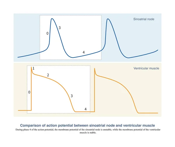
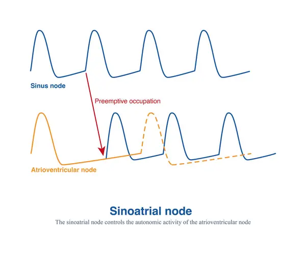
Stock image The autonomic frequency of the sinoatrial node is the fastest, and other secondary pacemakers are controlled through mechanisms of preemptive occupation and overspeed suppression.
Published: May.11, 2024 04:55:18
Author: asia11m
Views: 0
Downloads: 0
File type: image / jpg
File size: 7.29 MB
Orginal size: 10000 x 8364 px
Available sizes:
Level: beginner