photo free
Home Images Vectors Videos

Stock image When the left atrium is diseased, the depolarization time of the left atrium is prolonged, and the duration of the sinus P wave on the electrocardiogram increases by more than 120ms.

Stock image When the left atrium is diseased, the depolarization time of the left atrium is prolonged, and the duration of the sinus P wave on the electrocardiogram increases by more than 120ms.
Published: Apr.25, 2023 09:47:28
Author: asia11m
Views: 8
Downloads: 0
File type: image / jpg
File size: 8.16 MB
Orginal size: 10000 x 10091 px
Available sizes:
S
(495 x 500)
M
(991 x 1000)
L
(1982 x 2000)
XL
(10000 x 10091)
EL0
(10000 x 10091)
Level: beginner
Download Image
Image topics:

Similar stock images

stock image The depolarization time of the right atrium is prolonged, causing the co-depolarization time of the left and right atria to be prolonged, so the P wave amplitude increases.

The Depolarization Time Of The Right Atrium Is Prolonged, Causing The Co-depolarization Time Of The Left And Right Atria To Be Prolonged, So The P Wave Amplitude Increases.

9000 × 13726
stock image The patient, a 67 year old female, suddenly fainted during the onset of variant angina pectoris. ECG indicates polymorphic ventricular tachycardia, which was at risk of sudden death.

The Patient, A 67 Year Old Female, Suddenly Fainted During The Onset Of Variant Angina Pectoris. ECG Indicates Polymorphic Ventricular Tachycardia, Which Was At Risk Of Sudden Death.

10000 × 7893
stock image The electrocardiogram characteristic of right atrial abnormality is an increase in  P wave amplitude, with limb leads exceeding 2.5mm and chest leads exceeding 1.5mm.

The Electrocardiogram Characteristic Of Right Atrial Abnormality Is An Increase In P Wave Amplitude, With Limb Leads Exceeding 2.5mm And Chest Leads Exceeding 1.5mm.

10000 × 13345
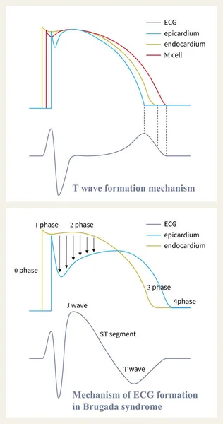
stock image The mechanism of Brugada syndrome is the difference of repolarization phase 1 and phase 2 between epicardium and endocardium, and the loss of epicardial plateau leads to ST segment elevation.

The Mechanism Of Brugada Syndrome Is The Difference Of Repolarization Phase 1 And Phase 2 Between Epicardium And Endocardium, And The Loss Of Epicardial Plateau Leads To ST Segment Elevation.

8000 × 15162
stock image Sometimes, evolution is found on the ECG of a patient with acute myocardial infarction, depending on the time point or lead of the ST-segment elevation.

Sometimes, Evolution Is Found On The ECG Of A Patient With Acute Myocardial Infarction, Depending On The Time Point Or Lead Of The ST-segment Elevation.

10000 × 8921
stock image In acute transmural myocardial ischemia, epicardial potassium outflow and myocardial interstitial potassium concentration increase, which is one of the reasons for the increase of ECG T wave.

In Acute Transmural Myocardial Ischemia, Epicardial Potassium Outflow And Myocardial Interstitial Potassium Concentration Increase, Which Is One Of The Reasons For The Increase Of ECG T Wave.

10000 × 12239
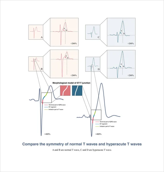
stock image It is worth noting that many electrocardiographic textbooks believe that hyperacute T waves are symmetrical. In fact, they are not symmetrical.

It Is Worth Noting That Many Electrocardiographic Textbooks Believe That Hyperacute T Waves Are Symmetrical. In Fact, They Are Not Symmetrical.

10000 × 10486
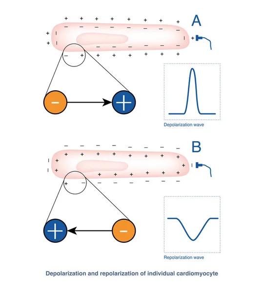
stock image When a single cardiomyocyte is depolarized, a positive wave is recorded towards the power supply; in the case of repolarization, electrodes in the same part record negative waves.

When A Single Cardiomyocyte Is Depolarized, A Positive Wave Is Recorded Towards The Power Supply; In The Case Of Repolarization, Electrodes In The Same Part Record Negative Waves.

10000 × 11087