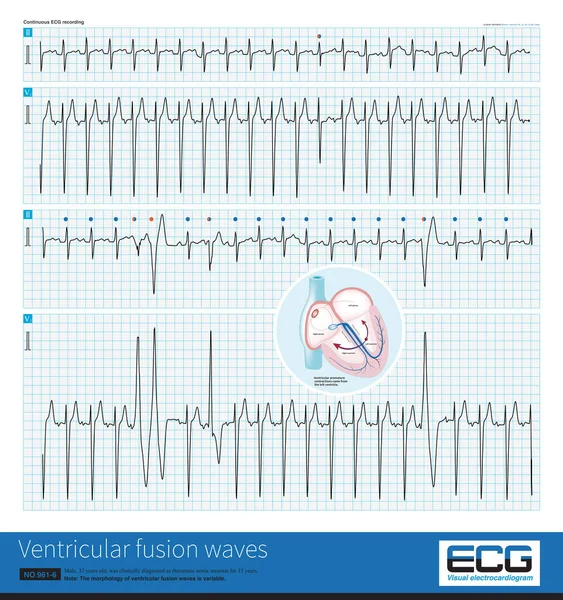
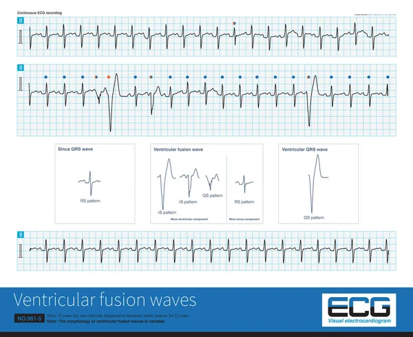
Stock image Ventricular premature contractions in late diastole can be combined with sinus impulses to excite the ventricles and produce ventricular fusion waves.

Published: Nov.07, 2022 14:58:44
Author: asia11m
Views: 16
Downloads: 0
File type: image / jpg
File size: 37.36 MB
Orginal size: 10000 x 10641 px
Available sizes:
Level: beginner