Home
Images
Vectors
Videos
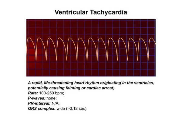
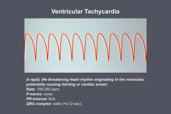
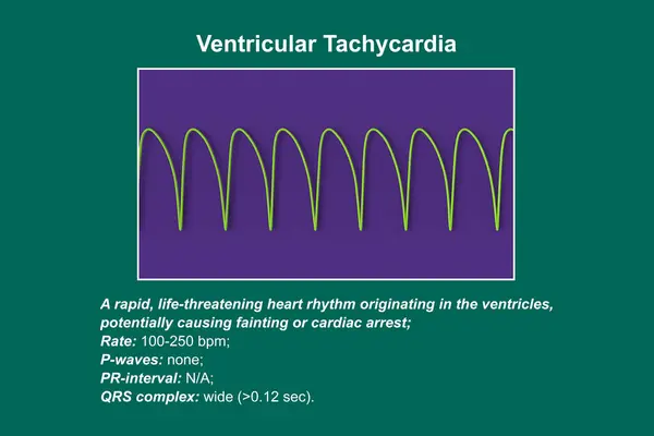
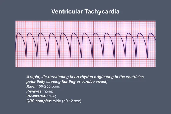
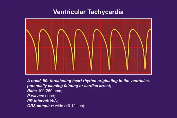
Stock image Ventricular tachycardia: A rapid heart rhythm originating in the ventricles, causes palpitations, dizziness, and life-threatening symptoms. ECG shows wide QRS complexes, 3D illustration.
Published:
Sep.26, 2023 14:24:42
Author:
katerynakon
Views:
1
Downloads:
0
File type:
image
/
jpg
File size:
8.07 MB
Orginal size:
9000
x
6000
px
Available sizes:
S
(500 x 333)
M
(1000 x 667)
L
(2000 x 1333)
XL
(9000 x 6000)
EL0
(9000 x 6000)
Level:
silver
Download Image
Image topics:
Illustration
Health
Pattern
Medicine
Healthcare
Medical
Electrical
3d
Biological
Cardiology
Heart
Monitor
Pulse
Disease
Conceptual
Condition
Node
Analysis
Graph
Beat
Poster
Rhythm
Heartbeat
Regular
Diagnostic
Tracing
Monitoring
Disorder
Impulse
Function
Conduction
Cardiac
Electrocardiogram
Abnormal
Contraction
Ecg
Waveforms
Tachycardia
Variability
Rhythms
Ventricular
Heart Rate
Heart Beat
Qrs
Anomalies
Abnormalities
Ventricular Tachycardia
Educatory
Sinus Tachycardia
Similar stock images
Ventricular Tachycardia: A Rapid Heart Rhythm Originating In The Ventricles, Causes Palpitations, Dizziness, And Life-threatening Symptoms. ECG Shows Wide QRS Complexes, 3D Illustration.
9000 × 6000
Ventricular Tachycardia: A Rapid Heart Rhythm Originating In The Ventricles, Causes Palpitations, Dizziness, And Life-threatening Symptoms. ECG Shows Wide QRS Complexes, 3D Illustration.
9000 × 6000
Ventricular Tachycardia: A Rapid Heart Rhythm Originating In The Ventricles, Causes Palpitations, Dizziness, And Life-threatening Symptoms. ECG Shows Wide QRS Complexes, 3D Illustration.
9000 × 6000
Ventricular Tachycardia: A Rapid Heart Rhythm Originating In The Ventricles, Causes Palpitations, Dizziness, And Life-threatening Symptoms. ECG Shows Wide QRS Complexes, 3D Illustration.
9000 × 6000
The QRS Wave Morphology Of Ventricular Tachycardia Can Be Consistent, Variable, Or Even Beat-to-beat Variant. The Latter Has Poorer Hemodynamics And Requires Urgent Treatment.
10000 × 4293
Male, 84 Years Old, Admitted To Hospital With Chest Pain For 1 Day. ECG Showed Acute Inferior And Posterior MI And Possibly Right MI. The Patient Died Of Ventricular Fibrillation The Next Day.
10000 × 4715
The Reentry Of Bundle Branch Reentrant Ventricular Tachycardia Occurs Between The Left Bundle Branch And The Right Bundle Branch. This Is A Malignant Ventricular Tachycardia.
10000 × 6444
Male, 48 Years Old, Clinically Diagnosed With Dilated Cardiomyopathy. Significant High-amplitude R Waves In Lead V5 Of The ECG Suggest Left Ventricular Hypertrophy.
10000 × 7291