Home
Images
Vectors
Videos
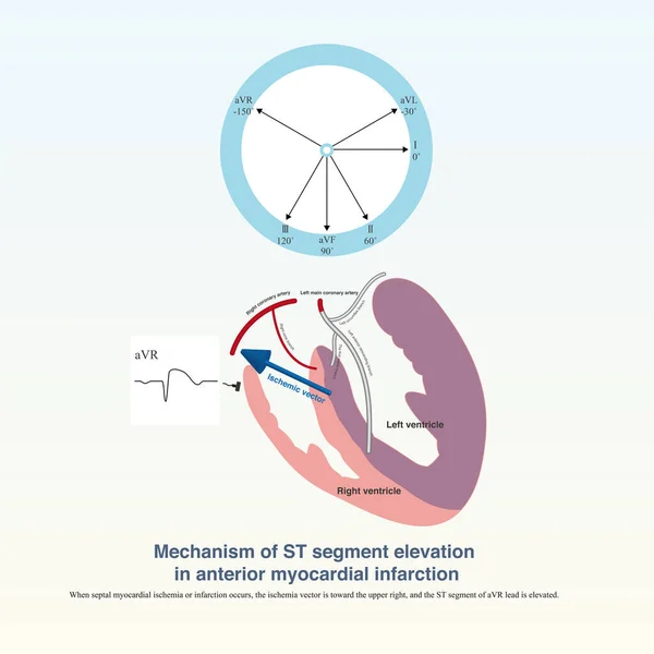
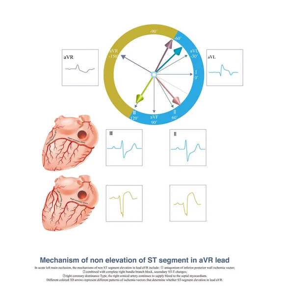
Stock image When acute left main trunk occlusion occurs, the ST segment of aVR lead may not be elevated, which is the result of the combined action of ischemic vector and corresponding vector.
Published:
Oct.04, 2022 06:22:30
Author:
asia11m
Views:
12
Downloads:
0
File type:
image
/
jpg
File size:
11.38 MB
Orginal size:
10000
x
10210
px
Available sizes:
S
(490 x 500)
M
(979 x 1000)
L
(1959 x 2000)
XL
(10000 x 10210)
EL0
(10000 x 10210)
Level:
beginner
Download Image
Image topics:
Background
Graphic
Illustration
Rate
Human
Health
Medicine
Medical
Wave
3d
Arrow
Cardiology
Heart
Pulse
Body
Disease
Science
Death
Anatomy
Circulation
Artery
Cardiogram
Cardiac
Ekg
Electrocardiogram
Ecg
Anatomical
Thrombus
Thrombosis
Heart Disease
Syncope
Cardiac Arrest
Coronary Artery
Sudden Death
Ischemic Heart Disease
Myocardial Ischemia
Acute Coronary Syndrome
ECG Monitoring
Coronary Spasm
St Segment
Ecg Education
Qrs Wave
Left Anterior Descending Artery
Ischemic Chest Pain
St Segment Elevation
Electric Alternation
Left Main Artery
Left Main Occlusion
Lateral Circulation
Similar stock images
In Case Of Acute Coronary Syndrome, If The ECG Shows Extensive ST Segment Depression, The Culprit Vessel May Be The Left Main Coronary Artery Or A Single Coronary Artery.
10000 × 6672
Sinus Node Electrogram Is A Technique To Record The Electrical Activity Of The Sinus Node In An Invasive Way. The Graph Contains The Depolarization Potential Of The Sinus Node And The Atrium.
10000 × 8072
In AMI, If The Amplitude Of ST Segment Elevation Is Less Than 1mm, It Is Called Slight ST Segment Elevation Myocardial Infarction, Which Is Easily Misdiagnosed As Early Repolarization
7000 × 9341
Generally, The Amplitude Of ST Segment Elevation Of AVR Lead 1mm Is Clinically Significant, And It Is Often Used For Criminal Vessel Locationand Prognosis Interpretation Of Acute Coronary Syndrome.
10000 × 9984
A 1-month-old Child Developed Atrioventricular Reentrant Tachycardia. When Tachycardia Occurred, Retrograde P Waves Can Be Detected Each QRS Complex.
10000 × 11672
Electrocardiography. ECG Or EKG Of A Heart With Normal Sinus Rhythm, ST Elevated Myocardial Infarction, Ishemia, ST Depression. Difference And Comparison. Vector Diagram For Education
4444 × 4443
Sometimes, Evolution Is Found On The ECG Of A Patient With Acute Myocardial Infarction, Depending On The Time Point Or Lead Of The ST-segment Elevation.
10000 × 8921
Acute Left Main Trunk Occlusion, Acute Left Anterior Descending Artery Occlusion And Acute Proximal Right Coronary Artery Occlusion Can All Cause ST Segment Elevation In AVR Lead.
10000 × 10000