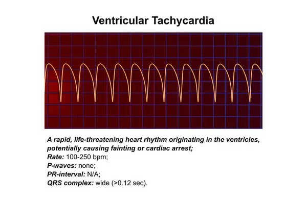
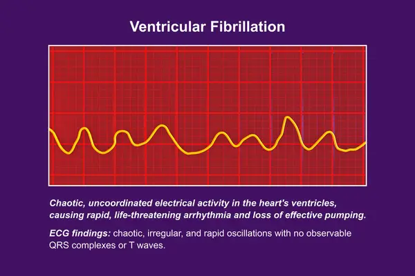
Stock image When the blood potassium concentration increases to a certain extent, atrial muscle paralysis accompanied by ventricular conduction disorder, ECG without sinus P wave accompanied by wide QRS waves.

Published: May.06, 2024 10:01:47
Author: asia11m
Views: 0
Downloads: 0
File type: image / jpg
File size: 7.2 MB
Orginal size: 10000 x 3728 px
Available sizes:
Level: beginner