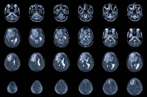
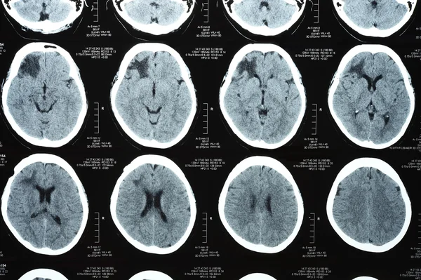
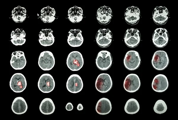
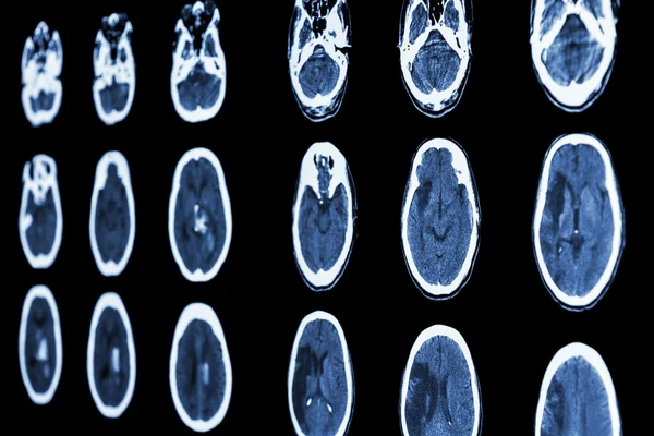
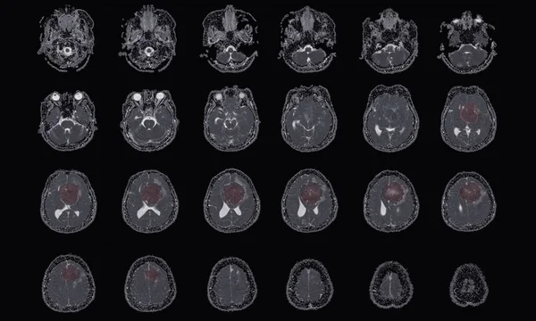
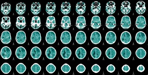
Stock image World Stroke Day,CT Scan Brain A man suffering from pulsating pain of headache. People medical healthcare and technology concept
Published: Sep.14, 2021 07:15:12
Author: Richmanphoto
Views: 6
Downloads: 1
File type: image / jpg
File size: 11.24 MB
Orginal size: 7008 x 3548 px
Available sizes:
Level: bronze