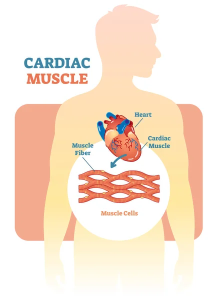
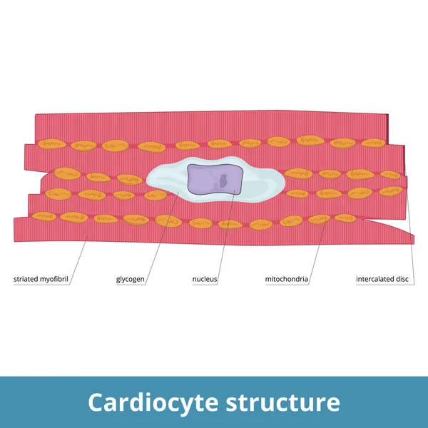
Stock vector Cardiocyte structure. Heart muscle cells and their elements include striated myofibril, glycogen, nucleus, and mitochondria. Intercalated disks as an adjacent part of them.
Published: Oct.14, 2022 13:44:40
Author: mortiara@gmail.com
Views: 3
Downloads: 1
File type: vector / eps
File size: 5.9 MB
Orginal size: 6250 x 6250 px
Available sizes:
Level: beginner