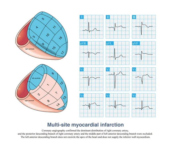
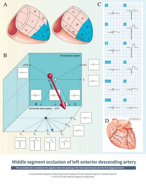
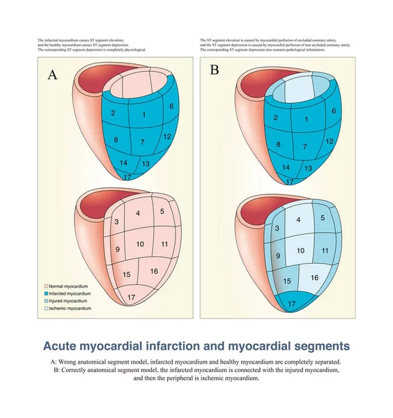
Stock image In imaging medicine, although the myocardium is divided into many segments, in anatomy, these myocardium are continuous rather than completely divided.
Published: Nov.15, 2022 10:17:51
Author: asia11m
Views: 28
Downloads: 0
File type: image / jpg
File size: 13.68 MB
Orginal size: 10000 x 10618 px
Available sizes:
Level: beginner