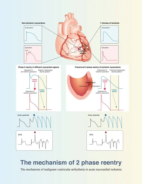
Stock image In the early stage of acute transmural myocardial ischemia, the mechanism of ventricular arrhythmia is mainly 2 phase reentry, including reentry between ischemic and non ischemic areas and reentry across the ventricular wall.
Published: Aug.22, 2022 09:53:07
Author: asia11m
Views: 22
Downloads: 0
File type: image / jpg
File size: 14.57 MB
Orginal size: 10000 x 12923 px
Available sizes:
Level: beginner
Similar stock images


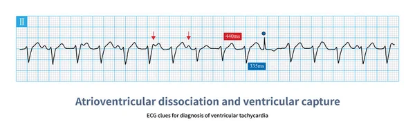
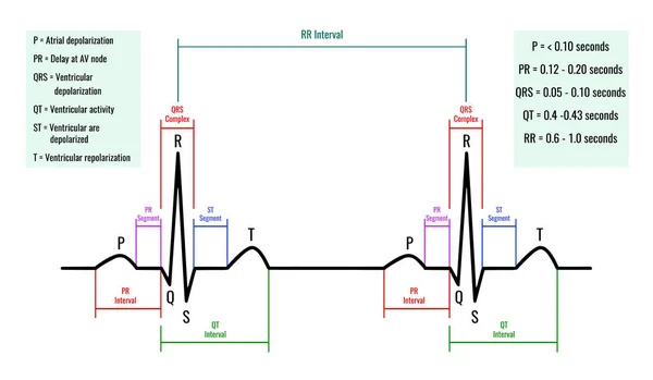
Illustration Of ECG Interpretation. ECG Of A Healthy Person. Useful For Educating Doctors And Nurses.
7087 × 4134