
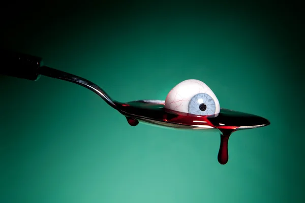
Stock image Bleeding Eyeball


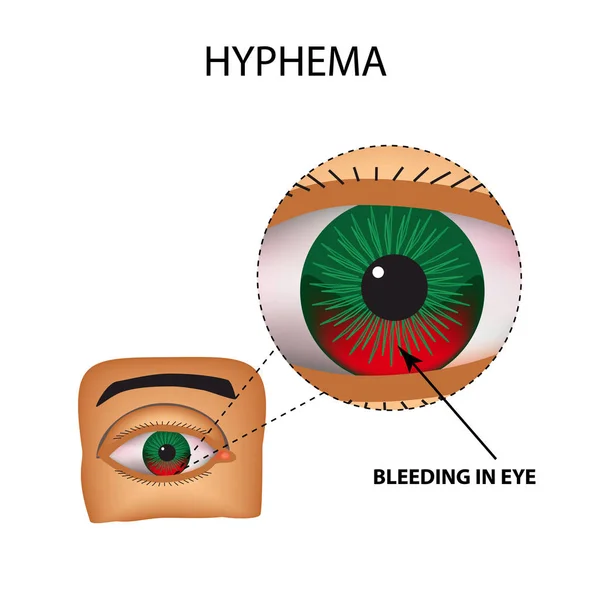
Hyphema. Anterior Eye Hemorrhage. The Structure Of The Eye. Infographics. Vector Illustration On Isolated Background
Vector, 1.27MB, 5000 × 5000 eps
Close-up Of A Woman's Green Eye With Burst Blood Vessels, Macrophotography
Image, 6.52MB, 6000 × 4000 jpg

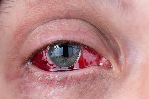
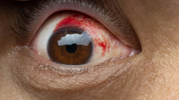
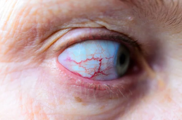
Blood In The Eye From A Subconjunctival Hemorrhage Usually Disappears Within A Week Or Two.Human Eye And Blood Close Up.
Image, 19.75MB, 6000 × 4000 jpg


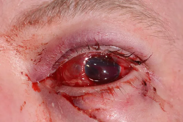
Blood In The Eye From A Subconjunctival Hemorrhage Usually Disappears Within A Week Or Two.Human Eye And Blood Close Up.
Image, 16.29MB, 6000 × 3375 jpg

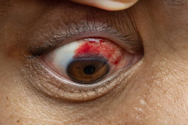
Blood In The Eye From A Subconjunctival Hemorrhage Usually Disappears Within A Week Or Two.Human Eye And Blood Close Up.
Image, 18.02MB, 5418 × 3612 jpg
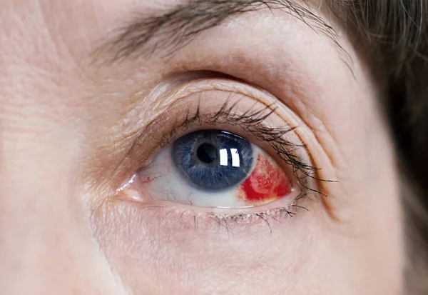

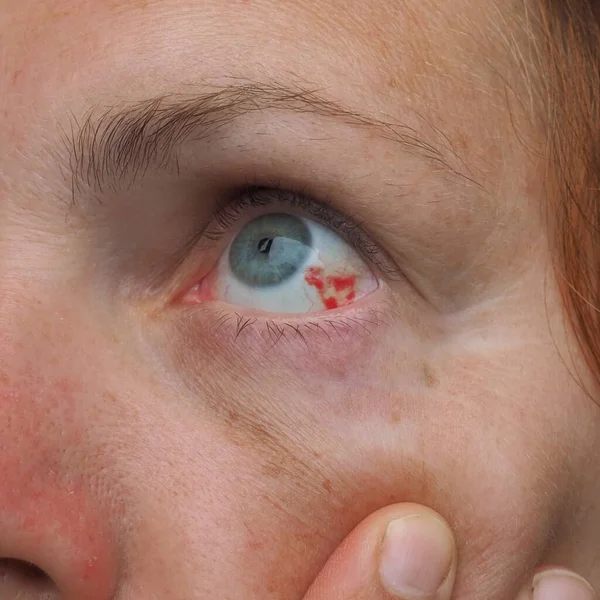
Close Up A A Caucasian Women's Blue Eye With A Subconjectival Hemorrhage, Or Broken Blood Vessel.
Image, 5.63MB, 3638 × 2425 jpg
The Vessel In The Eye Burst. Inflammation And Redness. The Structure Of The Eye. Infographics. Vector Illustration On Isolated Background
Vector, 1.27MB, 5000 × 5000 eps
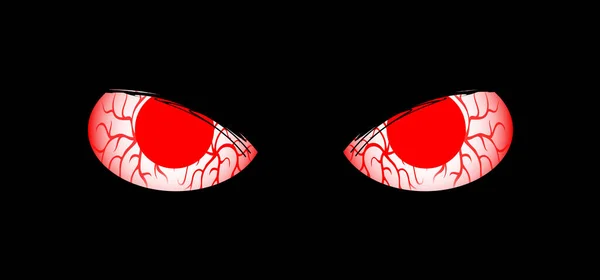
Ret Vein Eyes For Halloween Party. Eyes Of Cats Are In Darkness. Eyes Sparkle In The Dark, Animal Concept. Cartoon Eyes Icon. Kitty Silhouette
Image, 1.41MB, 5020 × 2345 jpg
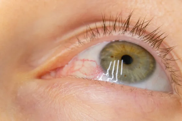
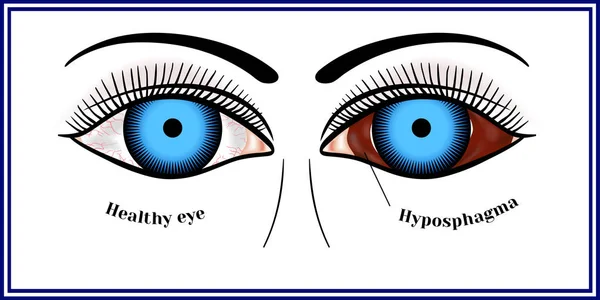
Subconjunctival Hemorrhage Or Subconjunctival Bleeding, Hyposhagmus. Bleeding Under The Conjunctiva. Damaged Blood Vessels. Blood Seeps Into The Space Between The Conjunctiva And The Sclera. Grey Eyes
Image, 4.5MB, 3456 × 3456 jpg
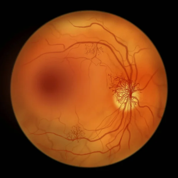
Proliferative Diabetic Retinopathy, Illustration Showing Neovascularization (formation Of New Vessels) In The Optic Disk And Other Sites. Fundoscopic Examination Of The Eye Retina In Diabetes Mellitus
Image, 2.86MB, 5000 × 5000 jpg
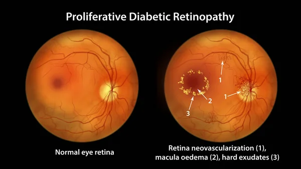
Proliferative Diabetic Retinopathy, Illustration Showing Neovascularization In The Disk And Other Sites, And Macula Edema. Fundoscopic Examination Of The Eye Retina In Diabetes Mellitus
Image, 2.9MB, 5000 × 5000 jpg

Proliferative Diabetic Retinopathy, Illustration Showing Neovascularization In The Disk And Macula Edema. Abnormal Finding On Fundoscopic Examination Of The Eye Retina In Diabetes Mellitus
Image, 2.85MB, 5000 × 5000 jpg
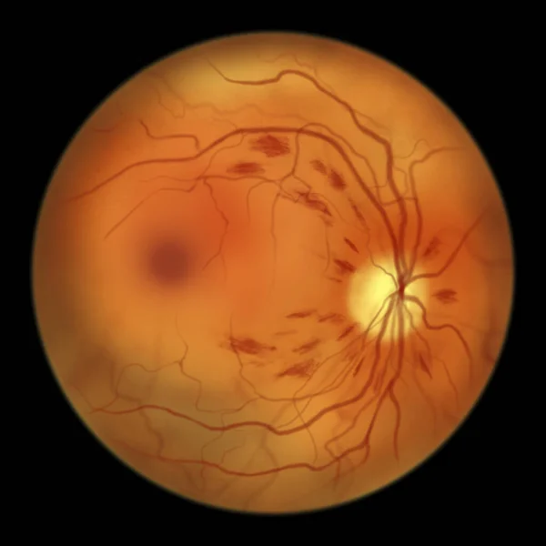
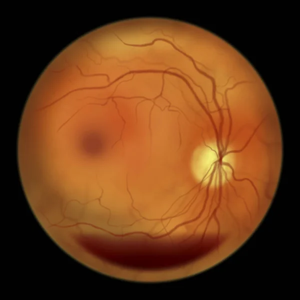
Non-proliferative Diabetic Retinopathy, Illustration Showing Flame-shaped And Splinter Retinal Haemorrhages, Ophthalmoscope View
Image, 2.96MB, 5000 × 5000 jpg
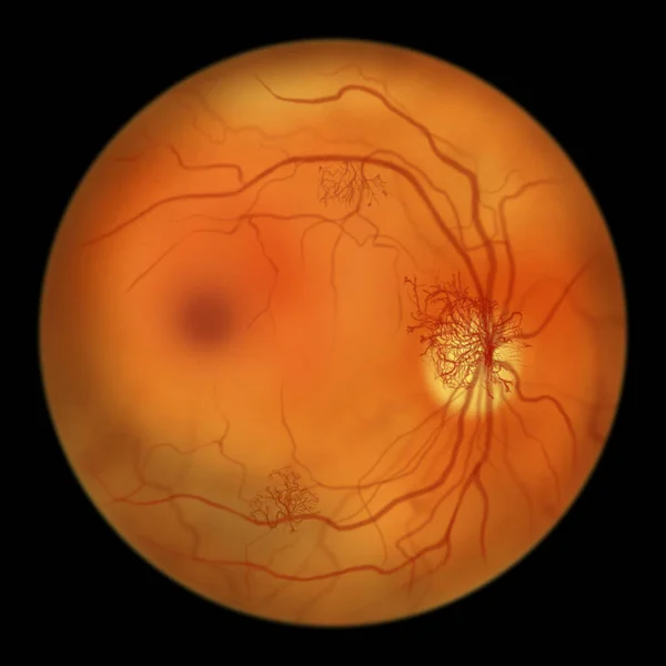
Proliferative Diabetic Retinopathy, Illustration Showing Neovascularization (formation Of New Vessels) In The Optic Disk. Fundoscopic Examination Of The Eye Retina In Diabetes Mellitus
Image, 2.81MB, 5000 × 5000 jpg
Diabetic Retinopathy, Illustration Shows Preretinal Haemorrhage As Horizontal Blood Level (boat-shaped Haemorrhage), Abnormal Finding On Fundoscopic Examination Of The Eye Retina In Diabetes Mellitus
Image, 2.69MB, 5000 × 5000 jpg
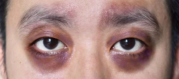
Raccon Eyes Or Periorbital Ecchymosis Or Panda Eye Sign In Southeast Asian Young Male Patient. It Is A Sign Of Basal Skull Fracture Or Subgaleal Hematoma.
Image, 3.93MB, 5481 × 3448 jpg
Southeast Asian, Chinese Young Man Hand Gripping, Hand Exercise Gripper.
Image, 2.71MB, 5593 × 2479 jpg

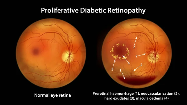
Proliferative Diabetic Retinopathy, Illustration Showing Neovascularization In The Disk And Other Sites, Macula Edema And Hard Exudates. Fundoscopic Examination Of The Eye Retina In Diabetes Mellitus
Image, 3.05MB, 5000 × 5000 jpg

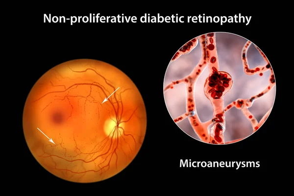
Non-proliferative Diabetic Retinopathy, 3D Illustration Showing Multiple Microaneurysms On The Eye Retina And Closeup View Of Microaneurysms, Microscopic Buldges In The Artery Walls Filled With Blood
Image, 11.11MB, 10431 × 6954 jpg
Non-proliferative Diabetic Retinopathy, 3D Illustration Showing Multiple Microaneurysms On The Eye Retina And Closeup View Of Microaneurysms, Microscopic Buldges In The Artery Walls Filled With Blood
Image, 10.46MB, 10431 × 6954 jpg
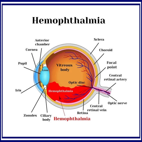
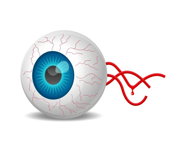
Hemophthalmia - The Presence Of Blood In The Vitreous Body (sometimes Called Hemorrhage In The Eyeball).
Vector, 0.65MB, 8333 × 8333 eps
Proliferative Diabetic Retinopathy, Illustration Showing Preretinal Haemorrhage As Horizontal Blood Level, Neovascularization In The Disk And Other Sites, Macula Edema And Hard Exudates
Image, 7.07MB, 11738 × 6603 jpg
Proliferative Diabetic Retinopathy, Illustration Showing Neovascularization In The Disk And Other Sites, Macula Edema And Hard Exudates. Fundoscopic Examination Of The Eye Retina In Diabetes Mellitus
Image, 6.95MB, 11738 × 6603 jpg

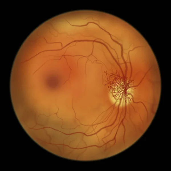
Proliferative Diabetic Retinopathy, Illustration Showing Neovascularization In The Optic Disk And Other Sites. Fundoscopic Examination Of The Eye Retina In Diabetes Mellitus, Fluorescein Angiography
Image, 6.07MB, 11738 × 6603 jpg


Diabetic Retinopathy, Ophthalmoscope View, Illustration Showing Accumulation Of Fatty Substances Leaked From Blocked Capillaries (yellow Patches), Haemorrhages (red Spots), Microaneurysms
Image, 4.38MB, 5000 × 5000 jpg

Non-proliferative Diabetic Retinopathy, 3D Illustration Showing Flame-shaped And Splinter Retinal Haemorrhages, Ophthalmoscope View
Image, 13.41MB, 5352 × 5352 jpg

Proliferative Diabetic Retinopathy, Illustration Showing Neovascularization In The Disk And Other Sites, And Macula Edema. Eye Retina In Diabetes Mellitus, Fluorescein Angiography
Image, 2.67MB, 5000 × 5000 jpg

Proliferative Diabetic Retinopathy, 3D Illustration Showing Preretinal Haemorrhage As Horizontal Blood Level, Neovascularization In The Disk And Other Sites, Macula Edema And Hard Exudates
Image, 22.98MB, 11738 × 6603 jpg

Preschooler Boy With Red Bursted Blood Vessels In Eye. Conjunctivitis, Trauma Of Eye, Inflammation, Infection, Allergy Or Intraocular Pressure Are Reason For Contacting An Ophthalmologist. Kids Health
Image, 2.69MB, 4000 × 2667 jpg
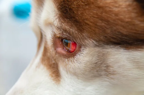
Eye Of A Dog With A Hemorrhage Resulting From A Blow To The Head. Siberian Husky Got Under The Car, A Traumatic Brain Injury.
Image, 11.18MB, 6016 × 4000 jpg

Hand Drawn Eyeball With Retinal Tear And Retinal Detachment. Disease Of The Organ Of Vision. Vector Illustration.
Vector, 0.35MB, 5679 × 3096 eps

A Patient With A Foot With Impaired Blood Circulation Has A Bandage On The Toe
Image, 12.57MB, 6000 × 4000 jpg

A Patient With A Foot With Impaired Blood Circulation Has A Bandage On The Toe
Image, 13.08MB, 6000 × 4000 jpg
Page 1 >> Next