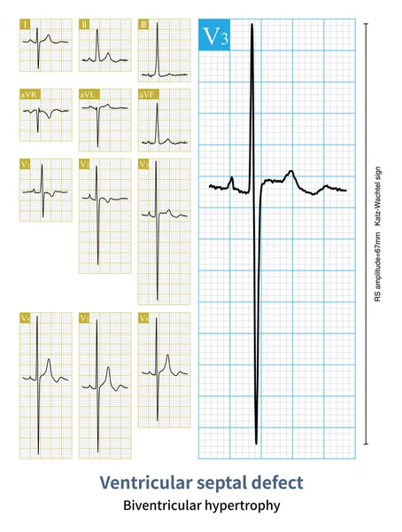
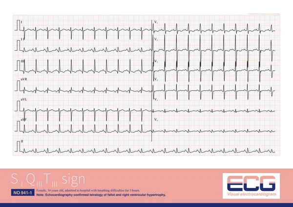
Stock image Male, 13 years old, clinically diagnosed with secundum atrial septal defect. Note that the QRS wave in lead V1 of the electrocardiogram has a qR shape, indicating right ventricular hypertrophy.

Published: Oct.09, 2023 07:37:37
Author: asia11m
Views: 3
Downloads: 1
File type: image / jpg
File size: 7.37 MB
Orginal size: 10000 x 7203 px
Available sizes:
Level: beginner