
Stock image Cardiac Ischemia

The Doctor Holds The Results Of An Examination Of An Elderly Woman Who Has Heart Problems. Cardiovascular Disease Concept, Ischemia And Angina Pectoris, Medical
Image, 1.6MB, 5400 × 3132 jpg

Mock Up Human Heart, Pills And Syringe On A Blue Background. Concept Of Heart Disease Treatment With Medication, Pharmacotherapy, Copy Space For Text, Antiplatelet Drugs
Image, 2.05MB, 5472 × 3648 jpg
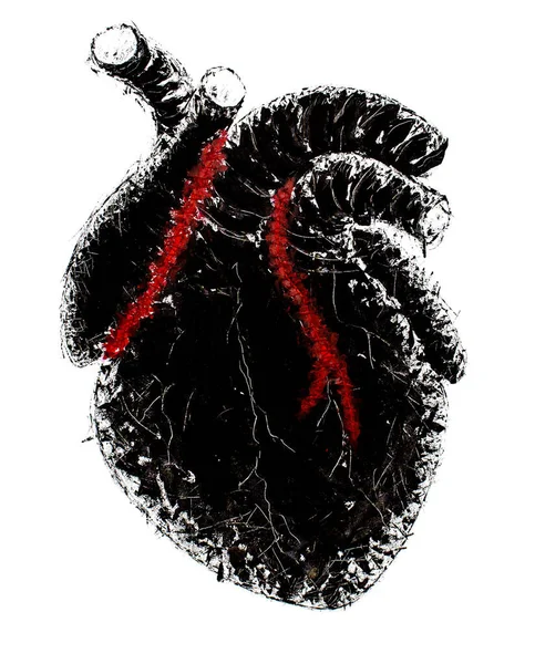
Stylized Drawing Of A Human Heart, Executed In Black And Red Colors On A White Background
Image, 4.16MB, 2696 × 3344 jpg

Anatomical Model Of The Organ Of The Heart On A Blue Background, Copy Space. The Concept Of Treatment And Diseases Of Myocardial Infarction And Heart Tachycardia, Cardiovascular Diseases
Image, 2.88MB, 4812 × 3528 jpg

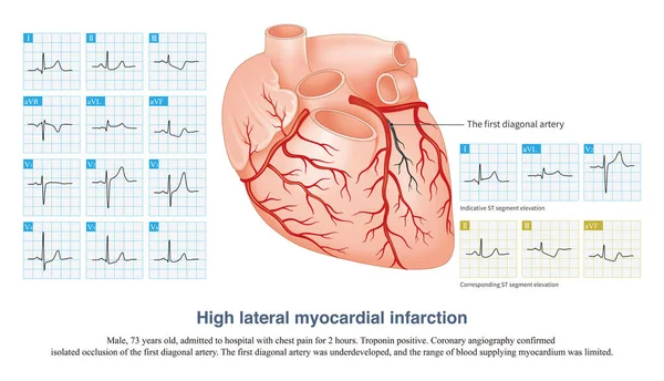
In Acute High Lateral Myocardial Infarction, There Is Indicative ST Segment Elevation In Leads I And AVL, And Corresponding ST Segment Depression In Leads II, III And AVF.
Image, 12.63MB, 10000 × 5739 jpg
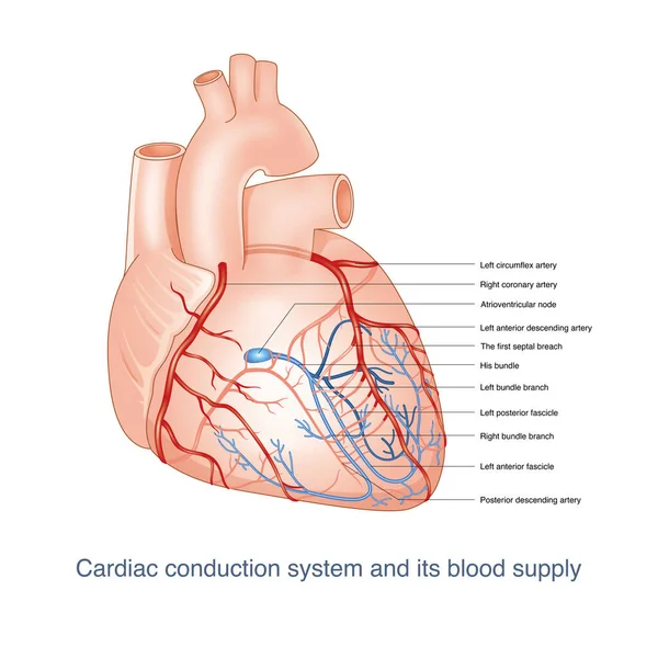
The Conduction System Of The Heart Is Supplied By The Branches Of The Coronary Artery. Once The Blood Vessels Are Blocked, It Can Cause Conduction Disorder. This Picture Is Suitable For Dark Background. This Picture Is Suitable For Light Background.
Image, 12.07MB, 10000 × 10000 jpg

3d Illustration Of The Heart In Internal Anatomy. Transparent On Blue Backlit Figure On White Background.
Image, 2.74MB, 5000 × 3750 jpg


General Practitioner, Cardiologist Or Paramedic Holding In Hand Tape With Recorded Electrocardiogram (EKG, ECG) Wave. Diagnostic Procedure To Detect Acute And Chronic Diseases Of Heart Using ECG
Image, 7.56MB, 6016 × 4000 jpg

Isometric Banner Augmented Reality In Medicine. Vector Illustration Projection Human Heart. Web Page Medical Clinic. Doctor Heart Disease Diagnosis. Modern Technologies Medical Treatment Patient
Vector, 1.39MB, 5076 × 4926 eps

Coronary Artery Spasm Causes Transmural Myocardial Ischemia, And ST Segment Elevation In ECG Has Localization Characteristics. Criminal Vessels Can Be Derived From ST Segment Elevation Leads In ECG.
Image, 11.66MB, 10000 × 7684 jpg

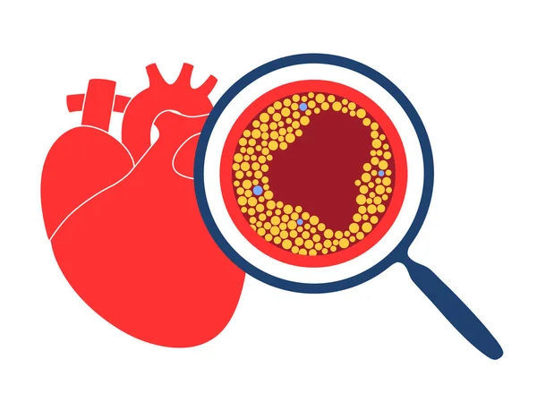
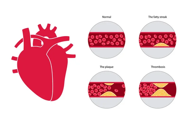
Coronary Heart Disease Symbol ( Ischemic Heart Disease , Myocardial Infraction ) Red Area At Coronary Artery ( Thrombus Occlude In Coronary Artery )
Vector, 3.59MB, 5000 × 5000 eps
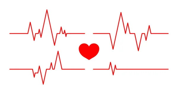
Heartbeat Line. Pulse And Cardiogram On Monitor. Icons Of Heart Beat. Ecg On Graph. Electrocardiogram With Healthy Rhythm, Cardio Attack, Ischemia, Infarction And Death. Symbol For Cardiac. Vector.
Vector, 0.26MB, 7509 × 4000 eps

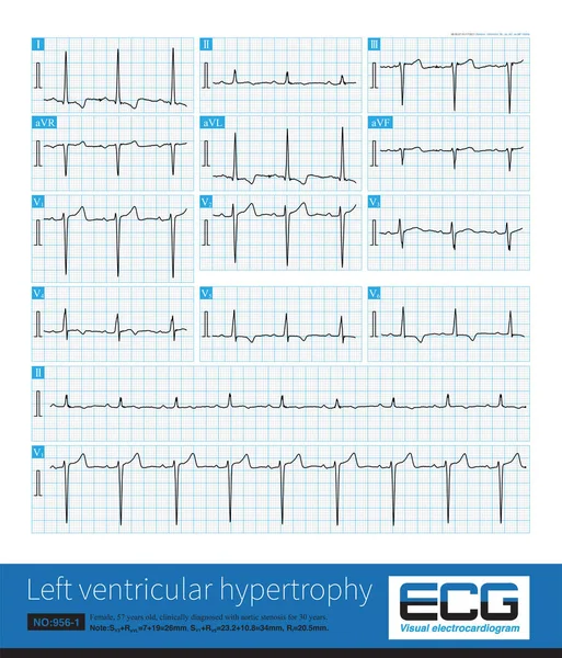
Sometimes, Because The QRS Axis Is In The Upper Left Quadrant, The High-amplitude R Wave Of Left Ventricular Hypertrophy Occurs In The Limb Leads, And Left Chest Leads Is Normal.
Image, 31.4MB, 10000 × 11694 jpg

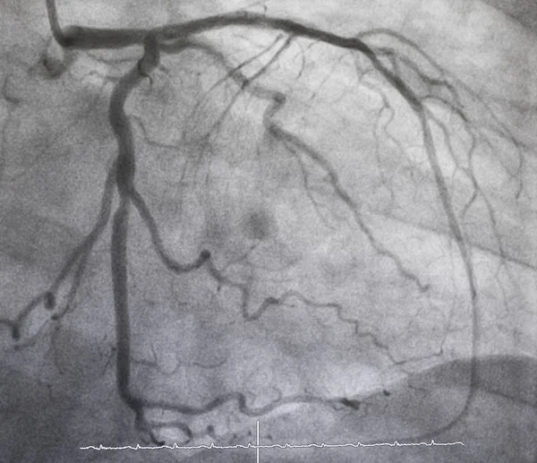
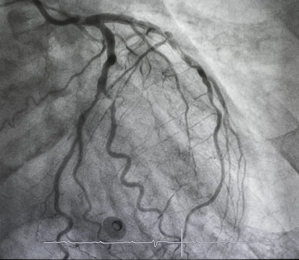
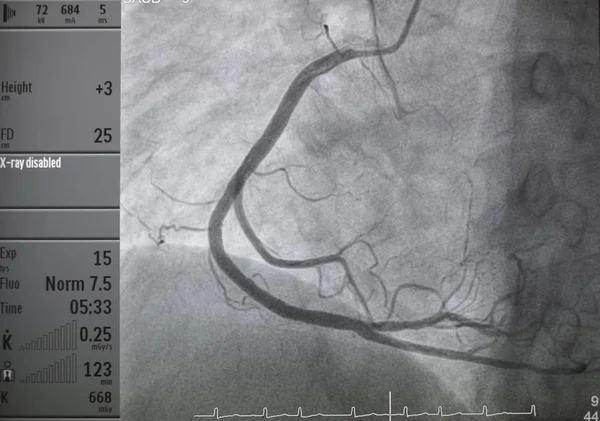
Coronary Angiography, Coronary Artery Disease. Medical X-ray Of Heart Disease. Healthcare And Medical Concept.
Image, 10.74MB, 6000 × 4063 jpg

3d Illustration Of Illustration Of A Blood Clot, Thrombus Or Embolus With Coagulated Red Blood Cells.
Image, 20.88MB, 6589 × 4942 jpg

Bidirectional Ventricular Tachycardia Is A Kind Of Malignant Arrhythmia. The Polarity Of QRS Main Wave Alternates From Beat To Beat, And It Is Easy To Degenerate Into Ventricular Fibrillation.
Image, 10.66MB, 10000 × 4450 jpg


Male, 60 Years Old, Clinically Diagnosed As Acute Extensive Anterior Wall Myocardial Infarction. The Patient Died Of Ventricular Fibrillation After Admission.
Image, 6.05MB, 10000 × 4656 jpg


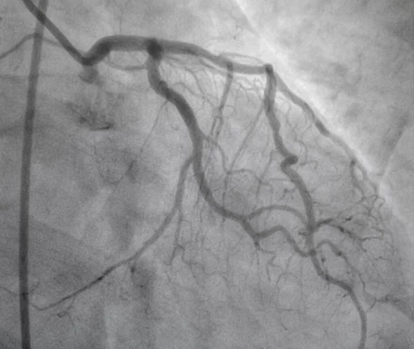
Coronary Angiography, Coronary Artery Disease. Medical X-ray Of Heart Disease. Healthcare And Medical Concept.
Image, 11.34MB, 6000 × 4000 jpg

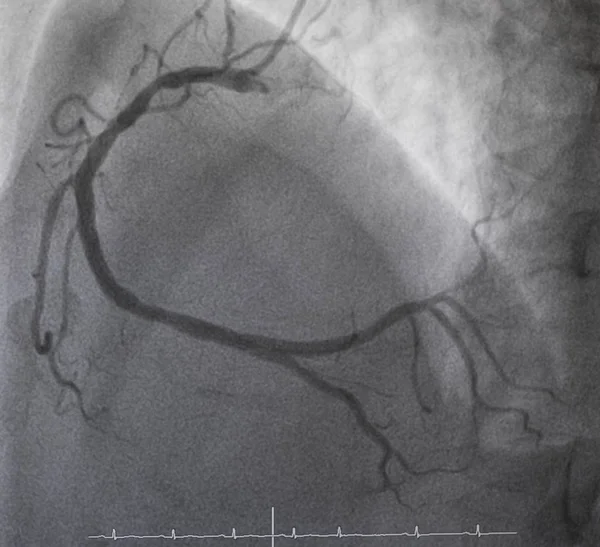
Coronary Angiography, Coronary Artery Disease. Medical X-ray Of Heart Disease. Healthcare And Medical Concept.
Image, 11.59MB, 6000 × 4000 jpg


3d Illustration Of Illustration Of A Blood Clot, Thrombus Or Embolus With Coagulated Red Blood Cells.
Image, 11.72MB, 6744 × 5058 jpg

Laboratory Medical Diagnostics, Tests For Heart And Cardiovascular Concept Photo. Doctor Or Laboratory Technician Holds In One Hand Laboratory Test Tube With Blood, In Other Hand - Figure Of Heart
Image, 7.43MB, 6000 × 4000 jpg

Coronary Angiography, Coronary Artery Disease. Medical X-ray Of Heart Disease. Healthcare And Medical Concept.
Image, 9.24MB, 6000 × 4087 jpg
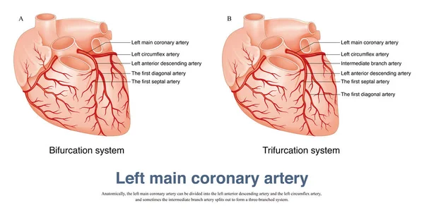
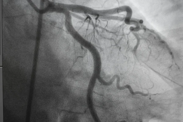
The Left Main Coronary Artery Can Be Divided Into The Left Anterior Descending Artery And The Left Circumflex Artery, And Sometimes The Intermediate Branch Artery.
Image, 7.94MB, 10000 × 5094 jpg
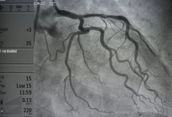
Coronary Angiogram , Medical X-ray For Heart Disease. Coronary Artery Disease.
Image, 11.11MB, 6000 × 5063 jpg

Male, 60 Years Old, Clinically Diagnosed As Acute Extensive Anterior Wall Myocardial Infarction. The Patient Died Of Ventricular Fibrillation After Admission.
Image, 10.58MB, 10000 × 6427 jpg


During The Onset Of Variant Angina Pectoris, ECG Is Divided Into Non Fusion Wave, Partial Fusion Wave And Complete Fusion Wave According To The Fusion Degree Of QRS Wave, ST Segment And T Wave.
Image, 7.94MB, 10000 × 6537 jpg
Page 1 >> Next