
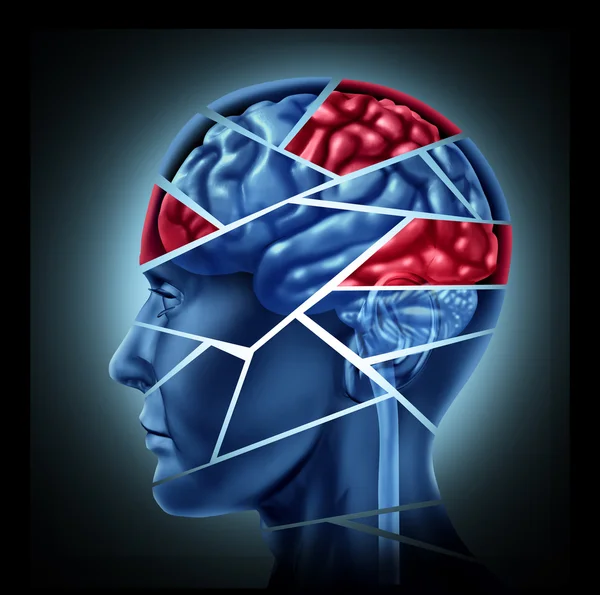
Stock image Degeneration Disease
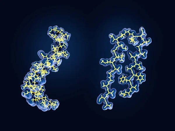
After Being Cleaved By The Gamma And Beta Secretases The Amyloid Beta Peptide, Which Has About 40 Amino Acid Residues, Leaves The Membrane, Changes Shape And Aggregates Into Long Fibrils. These Fibrils Form Dense Plaques On Nerve Cells.
Image, 2.61MB, 8000 × 6000 jpg


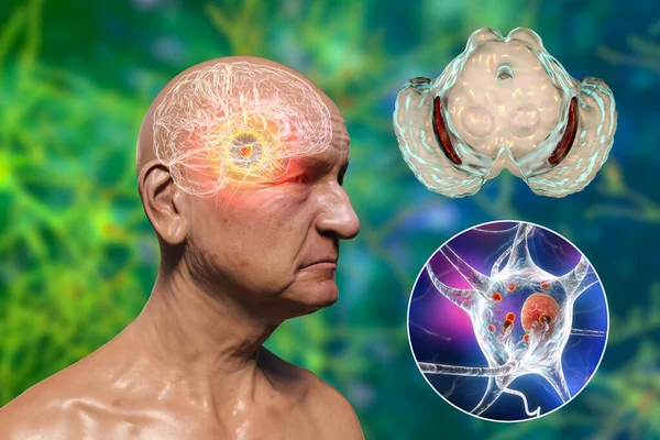
Infectious Etiology Of Dementia. Neuropsychiatric Sequelae Of Covid-19. Viruses Infecting Neurons And Progressive Impairment Of Brain Functions, Amyloid Plaques In Brain Tissues. 3D Illustration
Image, 14.68MB, 7200 × 4050 jpg

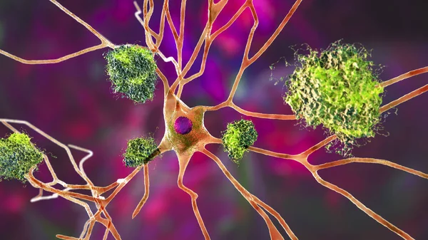
Neurons In Alzheimer's Disease. 3D Illustration Showing Amyloid Plaques In Brain Tissue, Neurofibrillary Tangles And Distruction Of Neuronal Networks
Image, 8.51MB, 7200 × 4050 jpg


An Old Man With Parkinson's Disease And Highlighted Black Substance Of The Midbrain. 3D Illustration Shows Decrease Of Substantia Volume And Accumulation Of Lewy Bodies In Its Dopaminergic Neurons
Image, 11.03MB, 6306 × 4203 jpg


Back View,Senior Woman Suffering From Neck Pain,disease Of Cervical Spondylosis,old Elderly Touching Nape Or Scruff,muscle Tension,injury In Occipital Bone,problems Of The Neck Joints,cervical Spine
Image, 6.04MB, 6485 × 4323 jpg

Intranuclear Neuronal Inclusions, 3D Illustration. Intranuclear Inclusions In Neurons Are Found In Different Neurodegenerative Diseases, Including Huntingon's Disease, Spinocerebellar Ataxia And Other
Image, 8.23MB, 7200 × 4050 jpg

Intranuclear Neuronal Inclusions, 3D Illustration. Intranuclear Inclusions In Neurons Are Found In Different Neurodegenerative Diseases, Including Huntingon's Disease, Spinocerebellar Ataxia And Other
Image, 9.43MB, 7200 × 4050 jpg


A Man In Blue Shirt Feeling Pain On His Back. Office Syndrome. Back Pain From Work. Herniated Nucleus Pulposus. Spine Pain. Spinal Degeneration.
Image, 3.03MB, 4896 × 1952 jpg

Neurons In Alzheimer's Disease. 3D Illustration Showing Amyloid Plaques In Brain Tissue, Neurofibrillary Tangles And Distruction Of Neuronal Networks
Image, 8.23MB, 7200 × 4050 jpg

Black Substance Of The Midbrain And Its Dopaminergic Neurons, 3D Illustration. Black Substance Regulates Movement And Reward, Its Degeneration Is A Key Step In Development Of Parkinson's Disease
Image, 2.54MB, 6000 × 4000 jpg


Neurons In Alzheimer's Disease. 3D Illustration Showing Amyloid Plaques In Brain Tissue, Neurofibrillary Tangles And Distruction Of Neuronal Networks
Image, 9.65MB, 7200 × 4050 jpg

Middle Aged Adult Woman Suffering From Arthritis Disease,Women Touching On Knee,Osteoarthritis
Image, 17.55MB, 8500 × 5674 jpg
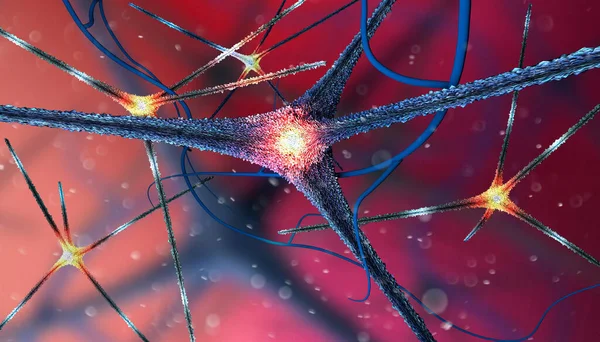
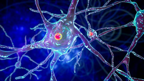
Microscopic View Of The Synapses. Brain Connections. Neurons And Synapses. Communication And Cerebral Stimulus. Neural Network Circuit, Degenerative Diseases, Parkinson, Alzheimer. 3d Render
Image, 10.63MB, 5511 × 3149 jpg

Man With Big Red Cracked Ear And Head, Symbolizing Tinnitus And Ear Problems.Male Head Stylized Profile. Photomontage With Dry Cracked Earth. Concept Symbolizing Tinnitus, Depression And Hearing Problems.
Image, 3.43MB, 3336 × 3660 jpg

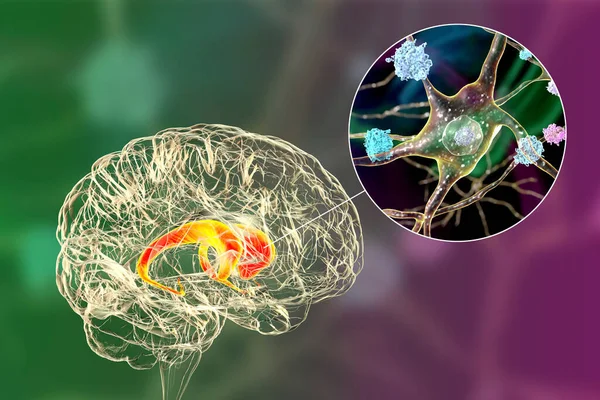
Dorsal Striatum, Caudate Nucleus And Putamen, Highlighted In The Brain Of A Person With Huntington's Disease And Close-up View Of Neuronal Degradation, Conceptual 3D Illustration
Image, 12.32MB, 7814 × 5210 jpg
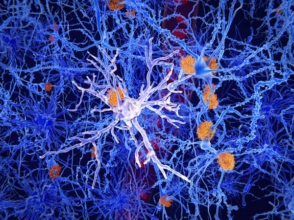
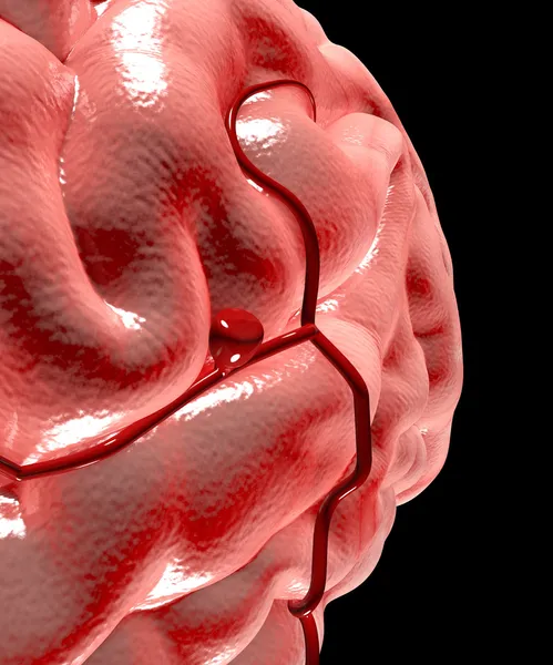
A Microglia Cell. It Plays An Important Role In The Pathogenesis Of Alzheimer's Disease
Image, 13.48MB, 8000 × 6000 jpg
Neuronal Inclusions In The Caudate Nucleus Of The Brain In Huntington's Disease, 3D Illustration. Inclusions Are Composed Of Mutated Huntingtin Protein, They Are Found In Nuclei, Axons And Dendrites
Image, 13.03MB, 7996 × 5331 jpg


Page 1 >> Next