
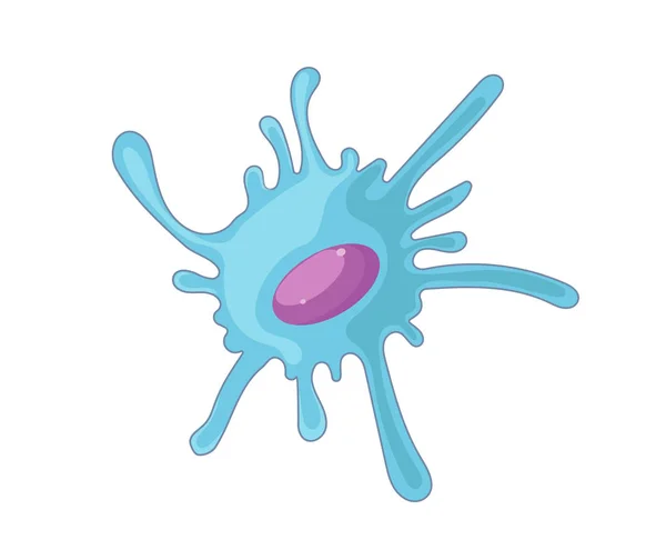
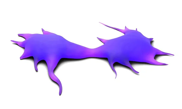
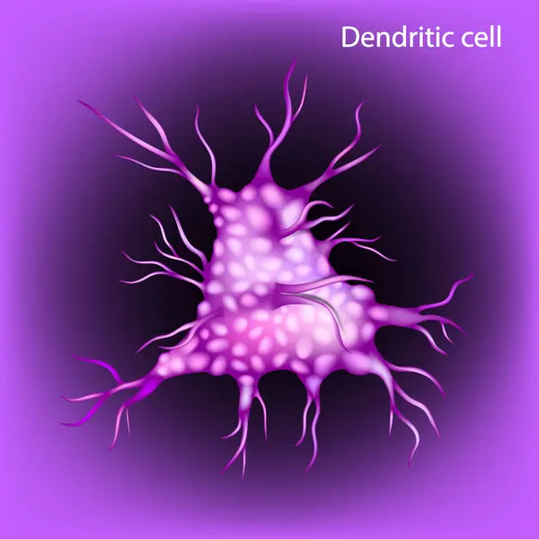
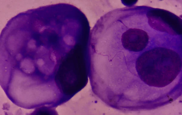
Stock image Dendritic Cell
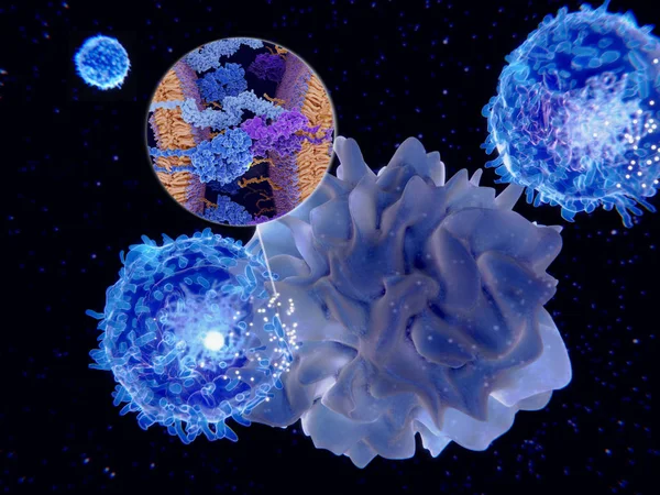
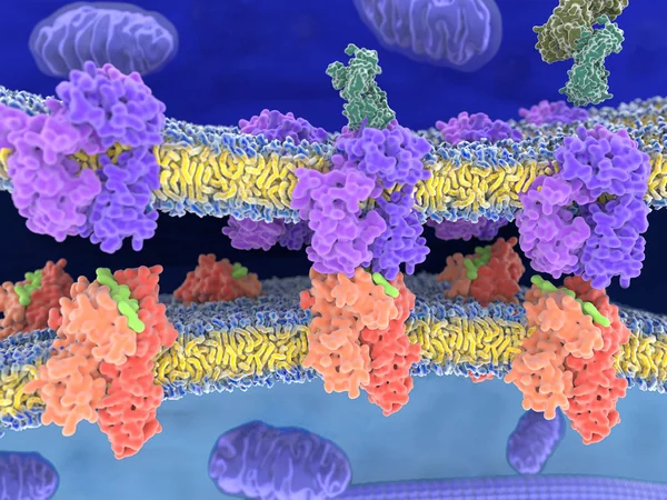
Dendritic Cells Present Antigens (green) To Lymphocytes Through Their Membran Bound MHC-molecules (violet). CD4 Molecules (light Blue) Bind To Other Portions Of The MHC, Strengthening The Interaction.
Image, 10.24MB, 8000 × 6000 jpg


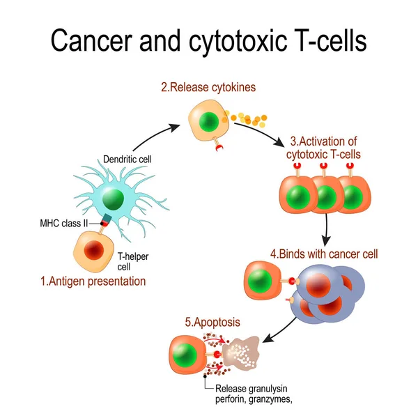
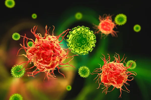
Cancer And Cytotoxic T-cells. T Lymphocyte Kills Cancer Cells. T-cell (immune Responses), Release The Perforin And Granzymes, And Attack Cancerous Cells. Through The Action Of Perforin, Granzymes Enter The Cytoplasm Of The Target Cell, And Lead To Ap
Vector, 1.01MB, 4050 × 4049 eps
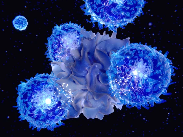
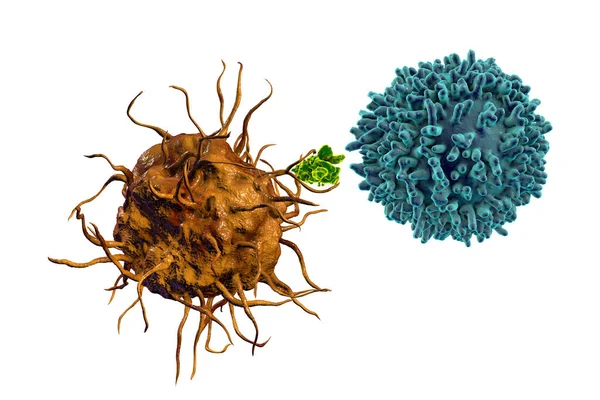
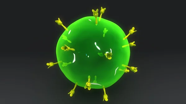
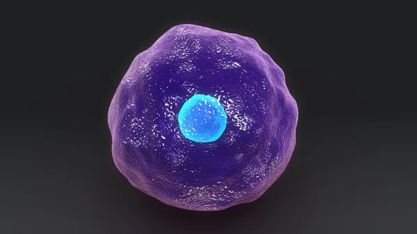
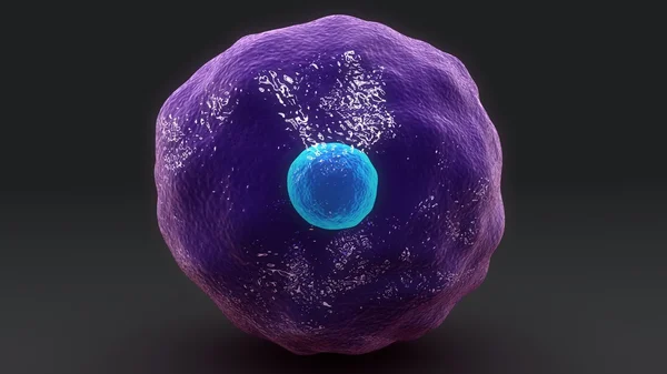
3d Computer Illustration Of A Dendritic Cell. They Areantigen-presenting Cells Of The Immune System. Their Main Function Is To Process Antigen Material And Present It On The Cell Surface To The T Cells Of The Immune System. They Are Messengers Betwe
Image, 5.2MB, 8000 × 6000 jpg

Interactions Of MHC-II With The T-cell Receptor And CD4 And B7-1 With CD-28 Activates T-cells While The Interactions Of P7-1 With CTLA-4 And PD-L1 With PD-1 Deactivates T-cells.
Image, 10.7MB, 8000 × 6000 jpg
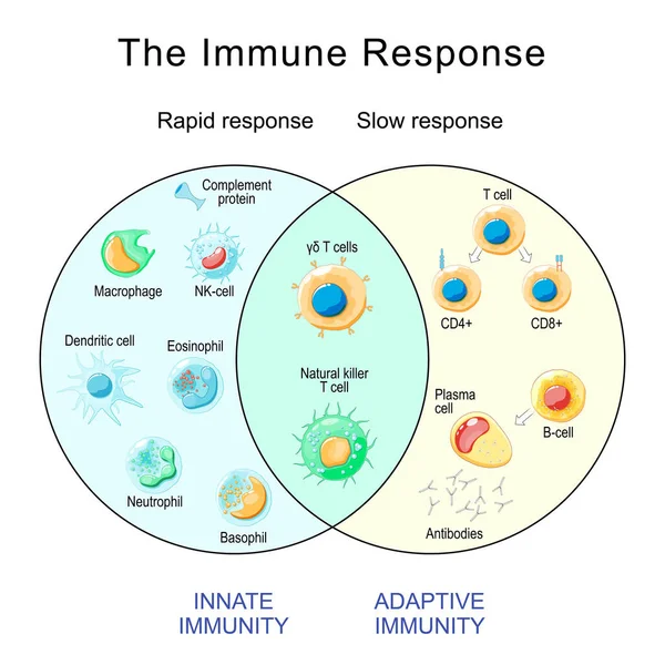
Immune Response. Rapid And Slow Response Of Adaptive And Innate Immunity And Antibody Activation. Cells Of The Immune System. Immunology Infographic. Vector Illustration
Vector, 2.28MB, 4444 × 4444 eps

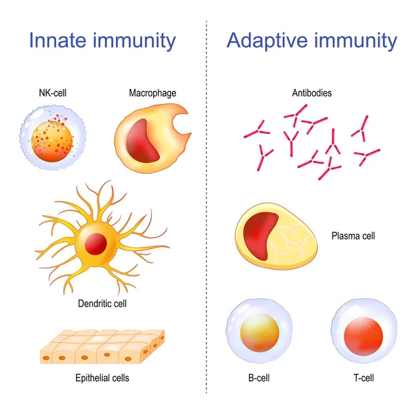
Adaptive Immunity: T-cell, Antibodies, Plasma Cell And B-cell. Innate Immunity: Macrophage, Dendritic, Epithelial, And NK Cells. Immunology Infographic. Vector Illustration
Vector, 14.03MB, 4444 × 4444 eps
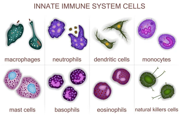
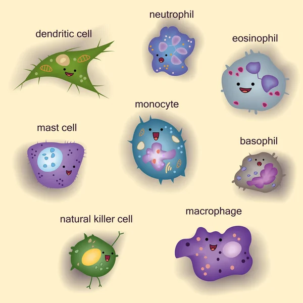
Set Of Innate Immune System Cells, Cartoon Cute Funny Vector Illustration
Vector, 7.73MB, 4792 × 4792 eps

IgA Immunity. Peyers Patch. Lymphoid Follicles Of The Small Intestine Generate IgA Immune Response With B Cells, T Cells, Dendritic Cells, And Plasma Cells Secreting IgA.
Vector, 6.98MB, 6250 × 5305 eps
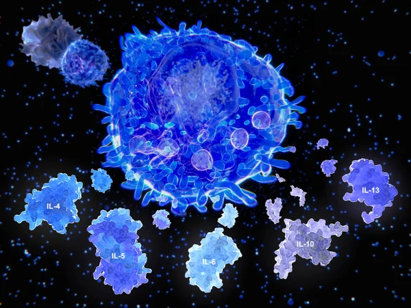
After Activation By An Antigen Presenting Cell, A T Helper Cell Segregates Several Cytokines.
Image, 13.65MB, 8000 × 6000 jpg

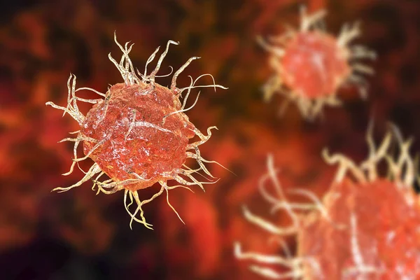
Dendritic Cells (DCs) Are Antigen-presenting Cells Also Known As Accessory Cells Of The Mammalian Immune System 3d Rendering
Image, 0.17MB, 2400 × 2000 jpg
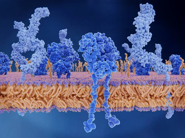
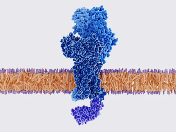
The T-cell Receptor Activates The Immune Response To Antigens In T-lymphocytes. T-cell Receptors (dark Blue), CD4 Molecules (light Blue), Glycolipids (orange). 3d Rendering. Illustration
Image, 3.11MB, 8000 × 6000 jpg
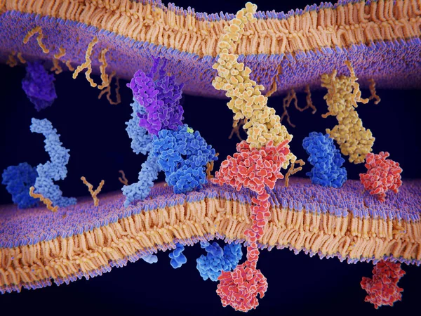
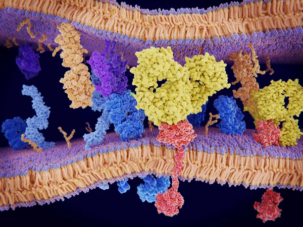
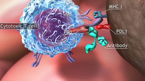
PD-1 (red) Extends From The Surface Of A T-cell And Interacts With The Ligand Protein PD-L1 (yellow) From A Antigen Presenting Cell. Although The T-cell Has Been Activated Through The Interaction Of A T-cell Receptor (blue) And A MHC Protein (viole
Image, 18.32MB, 8000 × 6000 jpg
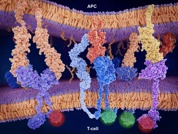
Interaction Of MHC-II (red) With The T-cell Receptor (blue) And CD4 (light Blue) And B7-1 (orange) With CD-28 (dark Blue) Activates T-cells While The Interaction Of P7-1 With CTLA-4 (violett) And PD-L1 (yellow) With PD-1 Deactivates T-cells
Image, 10.65MB, 8000 × 6000 jpg

Activation Of The Immune Response To An Antigene (green) Through The Complex Between A T-cell Receptor (dark Blue), An MHC II-antigen (violet) And A CD4 Protein (light Blue). 3d Rendering. Illustration
Image, 6.36MB, 8000 × 6000 jpg
Cancer Cells Express PD-L1 (orange) Proteins On Their Surface To Trick The Immune System. The Interaction Of PD-L1 With PD-1 Of T-cells Leads To A Down-regulation Of T-cells. The Antibody (yellow) Blocks This Interaction.
Image, 18.3MB, 8000 × 6000 jpg
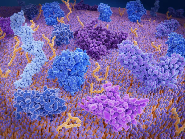
Immunologically Active Proteins On A T-cell. TCR (blue), CD-4 (light Blue), CD-28 (dark Blue), PD-1 (magenta), CTLA-4 (violet), Ca-channel (dark Violet). The T-cell Receptor, CD-4 And CD-28 Activate T-cells, While PD-1 And CTLA-4 Inhibit The Activat
Image, 10.2MB, 8000 × 6000 jpg
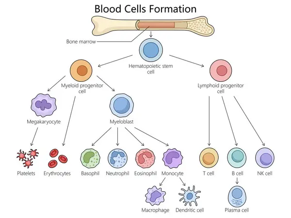
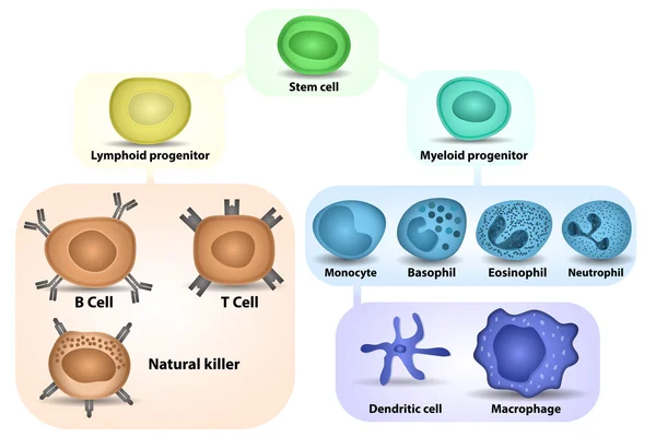
Human Hematopoiesis Blood Cell Formation From Bone Marrow, Hematopoietic Stem Cells Differentiation Structure Diagram Hand Drawn Schematic Raster Illustration. Medical Science Educational Illustration
Image, 3.18MB, 6000 × 4500 jpg

Human Hematopoiesis Blood Cell Formation From Bone Marrow, Hematopoietic Stem Cells Differentiation Structure Diagram Hand Drawn Schematic Vector Illustration. Medical Science Educational Illustration
Vector, 0.79MB, 5000 × 3750 eps

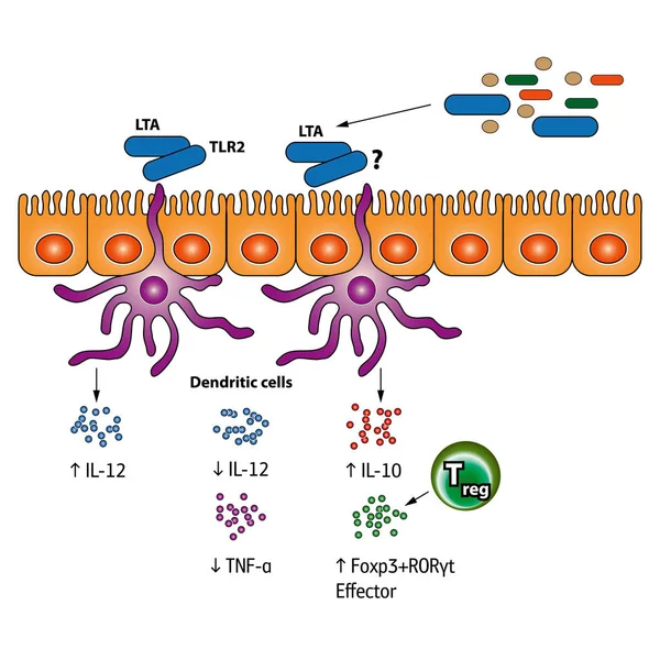
Maturation Dendritic Cells. Immature And Mature Dendritic Cells. Pathogens Cytokines
Vector, 2.14MB, 6000 × 3200 eps
3D Medical Illustration Cytotoxic T Cell And Helper T Cell Destroys The Stress-related Molecule
Image, 1.44MB, 3840 × 2160 jpg
T-cell Receptor In Complex With The MHC Class II-peptide Complex. The Antigen (light Green) Is A Peptide From A Tumor Cell, Bacteria Or Virus. Different Stages Of The Interaction. 3D-Rendering. Illustration
Image, 9.21MB, 8000 × 6000 jpg

T-cell Receptor In Complex With The MHC Class II-peptide Complex. The Antigen (light Green) Is A Peptide From A Tumor Cell, Bacteria Or Virus. Different Stages Of The Interaction. 3D-Rendering. Illustration
Image, 7.31MB, 8000 × 6000 jpg

Innate Immunity From Fever And Complement System (protein For Holes In The Plasma Membrane), To Macrophage, NK And Dendritic Cells. Adaptive Immunity From Antibodies And Plasma Cell To B-cell, T Helper, T-killer. Comparison And Difference
Vector, 2.86MB, 4444 × 4444 eps
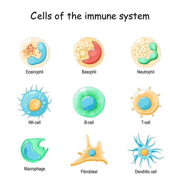
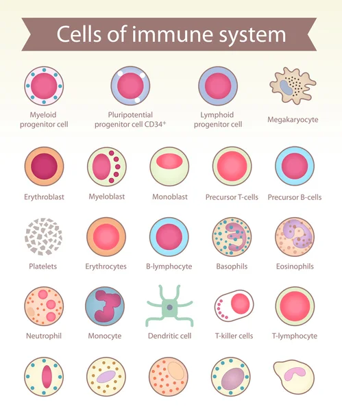
Cells Of The Immune System. White Blood Cells Or Leukocytes: Eosinophil, Neutrophil, Basophil, Macrophage, Fibroblast, And Dendritic Cell. Vector Diagram
Vector, 1.83MB, 4444 × 4444 eps
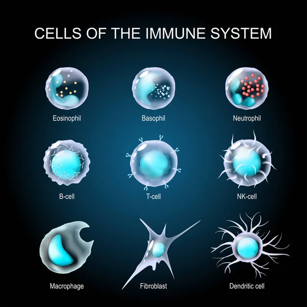
Cells Of The Immune System. White Blood Cells Or Leukocytes Eosinophil, Neutrophil, Basophil, Macrophage, Fibroblast, And Dendritic Cell. Set Of Transparent Realistic Cells On A Dark Background. Vector Illustration
Vector, 5.41MB, 4444 × 4444 eps
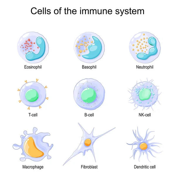
Cells Of The Immune System. White Blood Cells Or Leukocytes Eosinophil, Neutrophil, Basophil, Macrophage, Fibroblast, And Dendritic Cell. Vector Illustration
Vector, 1.73MB, 4444 × 4445 eps

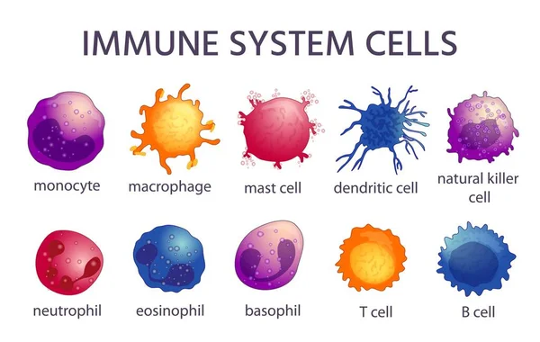
Immune System Cell Types. Cartoon Macrophage, Dendritic, Monocyte, Mast, B And T Cells. Adaptive And Innate Immunity, Lymphocyte Vector Set
Vector, 3.27MB, 6283 × 3979 eps
Page 1 >> Next