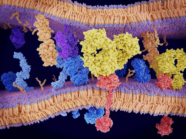
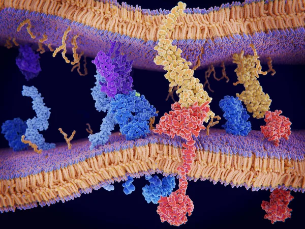
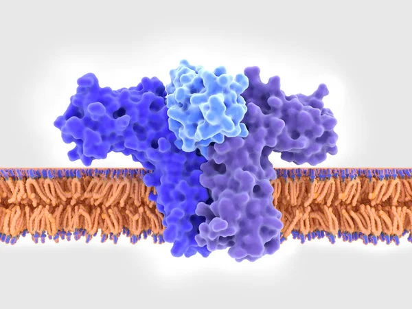
Stock image Activation of the immune response to an antigene (green) through the complex between a T-cell receptor (dark blue), an MHC II-antigen (violet) and a CD4 protein (light blue). 3d rendering. Illustration

Published: Mar.28, 2019 12:48:32
Author: animaxx3d
Views: 118
Downloads: 7
File type: image / jpg
File size: 6.36 MB
Orginal size: 8000 x 6000 px
Available sizes:
Level: bronze